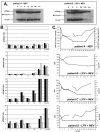
Monitoring processed, mature Human Immunodeficiency Virus type 1 particles immediately following treatment with a protease inhibitor-containing treatment regimen
- PMID: 15826302
- PMCID: PMC1087826
- DOI: 10.1186/1742-6405-2-2
Monitoring processed, mature Human Immunodeficiency Virus type 1 particles immediately following treatment with a protease inhibitor-containing treatment regimen
Abstract
Protease inhibitors (PIs) block HIV-1 maturation into an infectious virus particle by inhibiting the protease processing of gag and gag-pol precursor proteins. We have used a simple anti-HIV-1 p24 Western blot to monitor the processing of p55gag precursor into the mature p24 capsid immediately following the first dosage of a PI-containing treatment regimen. Evidence of PI activity was observed in plasma virus as early as 72 hours post treatment-initiation and was predictive of plasma viral RNA decrease at 4 weeks.
Figures


Similar articles
-
Maturation of human immunodeficiency virus particles assembled from the gag precursor protein requires in situ processing by gag-pol protease.AIDS Res Hum Retroviruses. 1991 May;7(5):475-83. doi: 10.1089/aid.1991.7.475. AIDS Res Hum Retroviruses. 1991. PMID: 1873082
-
Naturally occurring amino acid polymorphisms in human immunodeficiency virus type 1 (HIV-1) Gag p7(NC) and the C-cleavage site impact Gag-Pol processing by HIV-1 protease.Virology. 2002 Jan 5;292(1):137-49. doi: 10.1006/viro.2001.1184. Virology. 2002. PMID: 11878916
-
Morphogenic capabilities of human immunodeficiency virus type 1 gag and gag-pol proteins in insect cells.Virology. 1993 Mar;193(1):242-55. doi: 10.1006/viro.1993.1120. Virology. 1993. PMID: 8438569
-
Overexpression of the HIV-1 gag-pol polyprotein results in intracellular activation of HIV-1 protease and inhibition of assembly and budding of virus-like particles.Virology. 1993 Apr;193(2):661-71. doi: 10.1006/viro.1993.1174. Virology. 1993. PMID: 7681610
-
The virus-associated human immunodeficiency virus type 1 Gag-Pol carrying an active protease domain in the matrix region is severely defective both in autoprocessing and in trans processing of gag particles.Virology. 2004 Jan 20;318(2):534-41. doi: 10.1016/j.virol.2003.08.043. Virology. 2004. PMID: 14972522
References
-
- Arts EJ, Mak J, Kleiman L, Wainberg MA. DNA found in human immunodeficiency virus type 1 particles may not be required for infectivity. J Gen Virol. 1994;75:1605–1613. - PubMed
-
- Arts EJ, Mak J, Kleiman L, Wainberg MA. Mature reverse transcriptase (p66/p51) is responsible for low levels of viral DNA found in human immunodeficiency virus type 1 (HIV-1) Leukemia. 1994;8:S175–S178. - PubMed
-
- Coffin JM. Retroviridae: The viruses and thier replication. In: Fields BN, Knipe DM, Howley PM, editor. Fundamental Virology. 1996. pp. 763–843.
Grants and funding
LinkOut - more resources
Full Text Sources
Research Materials
Miscellaneous

