The neuronal ceroid lipofuscinosis Cln8 gene expression is developmentally regulated in mouse brain and up-regulated in the hippocampal kindling model of epilepsy
- PMID: 15826318
- PMCID: PMC1087490
- DOI: 10.1186/1471-2202-6-27
The neuronal ceroid lipofuscinosis Cln8 gene expression is developmentally regulated in mouse brain and up-regulated in the hippocampal kindling model of epilepsy
Abstract
Background: The neuronal ceroid lipofuscinoses (NCLs) are a group of inherited neurodegenerative disorders characterized by accumulation of autofluorescent material in many tissues, especially in neurons. Mutations in the CLN8 gene, encoding an endoplasmic reticulum (ER) transmembrane protein of unknown function, underlie NCL phenotypes in humans and mice. The human phenotype is characterized by epilepsy, progressive psychomotor deterioration and visual loss, while motor neuron degeneration (mnd) mice with a Cln8 mutation show progressive motor neuron dysfunction and retinal degeneration.
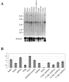
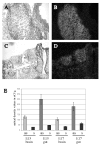
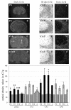
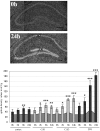
Results: We investigated spatial and temporal expression of Cln8 messenger ribonucleic acid (mRNA) using in situ hybridization, reverse transcriptase polymerase chain reaction (RT-PCR) and northern blotting. Cln8 is ubiquitously expressed at low levels in embryonic and adult tissues. In prenatal embryos Cln8 is most prominently expressed in the developing gastrointestinal tract, dorsal root ganglia (DRG) and brain. In postnatal brain the highest expression is in the cortex and hippocampus. Expression of Cln8 mRNA in the central nervous system (CNS) was also analyzed in the hippocampal electrical kindling model of epilepsy, in which Cln8 expression was rapidly up-regulated in hippocampal pyramidal and granular neurons.
Conclusion: Expression of Cln8 in the developing and mature brain suggests roles for Cln8 in maturation, differentiation and supporting the survival of different neuronal populations. The relevance of Cln8 up-regulation in hippocampal neurons of kindled mice should be further explored.
Figures


Similar articles
-
Localization of wild-type and mutant neuronal ceroid lipofuscinosis CLN8 proteins in non-neuronal and neuronal cells.J Neurosci Res. 2004 Jun 15;76(6):862-71. doi: 10.1002/jnr.20133. J Neurosci Res. 2004. PMID: 15160397
-
Different early ER-stress responses in the CLN8(mnd) mouse model of neuronal ceroid lipofuscinosis.Neurosci Lett. 2011 Jan 25;488(3):258-62. doi: 10.1016/j.neulet.2010.11.041. Epub 2010 Nov 19. Neurosci Lett. 2011. PMID: 21094208
-
The neuronal ceroid lipofuscinoses in human EPMR and mnd mutant mice are associated with mutations in CLN8.Nat Genet. 1999 Oct;23(2):233-6. doi: 10.1038/13868. Nat Genet. 1999. PMID: 10508524
-
Role of neurotrophin signalling in the differentiation of neurons from dorsal root ganglia and sympathetic ganglia.Cell Tissue Res. 2009 Jun;336(3):349-84. doi: 10.1007/s00441-009-0784-z. Epub 2009 Apr 23. Cell Tissue Res. 2009. PMID: 19387688 Review.
-
Northern epilepsy, a new member of the NCL family.Neurol Sci. 2000;21(3 Suppl):S43-7. doi: 10.1007/s100720070039. Neurol Sci. 2000. PMID: 11073227 Review.
Cited by
-
Caregiver-child interaction and early childhood development among preschool children in rural China: the possible role of blood epigenome-wide DNA methylation.BMC Genomics. 2025 Apr 1;26(1):329. doi: 10.1186/s12864-025-11406-2. BMC Genomics. 2025. PMID: 40170191 Free PMC article.
-
Role of endoplasmic reticulum stress in the amygdaloid kindling model of rats.Neurochem Res. 2011 Oct;36(10):1834-9. doi: 10.1007/s11064-011-0501-7. Epub 2011 May 22. Neurochem Res. 2011. PMID: 21604154
-
Analysis of NCL Proteins from an Evolutionary Standpoint.Curr Genomics. 2008 Apr;9(2):115-36. doi: 10.2174/138920208784139573. Curr Genomics. 2008. PMID: 19440452 Free PMC article.
-
The involvement of Purkinje cells in progressive myoclonic epilepsy: Focus on neuronal ceroid lipofuscinosis.Neurobiol Dis. 2023 Sep;185:106258. doi: 10.1016/j.nbd.2023.106258. Epub 2023 Aug 11. Neurobiol Dis. 2023. PMID: 37573956 Free PMC article. Review.
-
Sex-split analysis of pathology and motor-behavioral outcomes in a mouse model of CLN8-Batten disease reveals an increased disease burden and trajectory in female Cln8mnd mice.Orphanet J Rare Dis. 2022 Nov 11;17(1):411. doi: 10.1186/s13023-022-02564-7. Orphanet J Rare Dis. 2022. PMID: 36369162 Free PMC article.
References
-
- Santavuori P. Neuronal ceroid-lipofuscinoses in childhood. Brain Dev. 1988;10:80–83. - PubMed
-
- Haltia M. The neuronal ceroid-lipofuscinoses. J Neuropathol Exp Neurol. 2003;62:1–13. - PubMed
-
- Ranta S, Zhang Y, Ross B, Lonka L, Takkunen E, Messer A, Sharp J, Wheeler R, Kusumi K, Mole S, Liu W, Soares MB, Bonaldo MF, Hirvasniemi A, de la Chapelle A, Gilliam TC, Lehesjoki AE. The neuronal ceroid lipofuscinoses in human EPMR and mnd mutant mice are associated with mutations in CLN8. Nat Genet. 1999;23:233–236. doi: 10.1038/13868. - DOI - PubMed
-
- Messer A, Flaherty L. Autosomal dominance in a late-onset motor neuron disease in the mouse. J Neurogenet. 1986;3:345–355. - PubMed
Publication types
MeSH terms
Substances
LinkOut - more resources
Full Text Sources
Medical
Molecular Biology Databases
Miscellaneous

