

Comment
doi: 10.1073/pnas.0501516102.
Epub 2005 Apr 12.
Organization of synaptic myonuclei by Syne proteins and their role during the formation of the nerve-muscle synapse
Affiliations
- PMID: 15827115
- PMCID: PMC556304
- DOI: 10.1073/pnas.0501516102
Item in Clipboard
Comment
Organization of synaptic myonuclei by Syne proteins and their role during the formation of the nerve-muscle synapse
Proc Natl Acad Sci U S A.
.
No abstract available
Figures
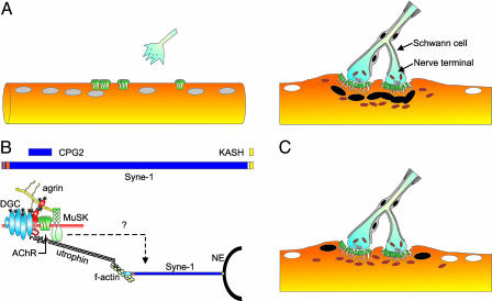
Development of the NMJ and the role of Syne-1. (A Left) Motor axons approach muscle fibers with myonuclei (gray) expressing high levels of clustered AChRs (green). (A Right) Mature NMJ with the presynaptic terminal containing aggregates of synaptic vesicles (white) and many mitochondria (orange). Nerve terminals are wrapped by Schwann cells. At the postsynaptic apparatus, AChRs (green) are concentrated at the crest of postsynaptic folds and myonuclei (black) and mitochondria accumulate. Extrasynaptic myonuclei (white) are transcriptionally distinct form subsynaptic ones. (B Upper) Structure of mouse Syne-1, the brain-specific alternative splice variant CPG2, and the dominant-negative form encoding the KASH domain used by Grady et al. (3). Red, calponin-type domains; blue, spectrin-like repeats; yellow, KASH domain. (B Lower) At the NMJ, agrin binds to the dystrophin-glycoprotein complex (DGC) via dystroglycan (red), activates MuSK, and aggregates AChRs. Utrophin binds to the DGC and the f-actin cytoskeleton. Syne-1 binds to f-actin and links it with the nuclear envelope (NE) via its KASH domain. Scheme is modified from ref. . (C) Mice overexpressing the KASH domain of Syne-1 lack subsynaptic myonuclei, and mitochondria are moving to the vicinity of the postsynaptic membrane. Gene expression in the perisynaptic (black) and extrasynaptic (white) myonuclei is as in wild-type mice.
Comment on
-
Syne proteins anchor muscle nuclei at the neuromuscular junction.Proc Natl Acad Sci U S A. 2005 Mar 22;102(12):4359-64. doi: 10.1073/pnas.0500711102. Epub 2005 Mar 4. Proc Natl Acad Sci U S A. 2005. PMID: 15749817 Free PMC article.
References
Publication types
MeSH terms
Substances
LinkOut - more resources
Full Text Sources
