. 2005 Apr 19;102(16):5826-31.
doi: 10.1073/pnas.0501316102. Epub 2005 Apr 12.

Late-onset motoneuron disease caused by a functionally modified AMPA receptor subunit

Affiliations

Late-onset motoneuron disease caused by a functionally modified AMPA receptor subunit

Rohini Kuner et al. Proc Natl Acad Sci U S A. .

Abstract

Amyotrophic lateral sclerosis (ALS) is a devastating disorder of the central nervous system in middle and old age that leads to progressive loss of spinal motoneurons. Transgenic mice overexpressing mutated human Cu(2+)/Zn(2+) superoxide dismutase 1 (SOD1) reproduce clinical features of the familial form of ALS. However, changes in SOD1 activity do not correlate with severity of motor decline in sporadic cases, indicating that targets unrelated to superoxide metabolism contribute to the pathogenesis of the disease. We show here that transgenic expression in mice of GluR-B(N)-containing L-alpha-amino-3-hydroxy-5-methylisoxazole-4-proprionate (AMPA) receptors with increased Ca(2+) permeability leads to late-onset degeneration of neurons in the spinal cord and decline of motor functions. Neuronal death progresses over the entire lifespan but manifests clinically in late adulthood, resembling the course of a slow neurodegenerative disorder. Additional transgenic expression of mutated human SOD1 accelerates disease progression, aggravates the severity of motor decline, and decreases survival. These observations link persistently elevated Ca(2+) influx through AMPA channels with progressive motor decline and late-onset degeneration of spinal motoneurons, indicating that functionally altered AMPA channels may be causally related to pathogenesis of sporadic ALS in humans.


Figures

Fig. 1.
Fig. 1.
Growth (a), locomotor activity (be), and gait patterns (f) in GluR-B(N) and WT mice between 19 and 83 weeks of age. Growth (a) was monitored by measurements of body weight (in grams) in GluR-B(N) (n = 9) and WT (n = 15) mice. ANOVA showed that although growth in GluR-B(N) and WT mice depended on age [F19–83(16,222) = 3.35, P < 0.001], it was significantly lower in GluR-B(N) than in WT mice [F19–83(1,222) = 355.73, P < 0.001]. Locomotor activity was monitored by means of horizontal activity (b), total distance (c), and average speed (d) in independent groups of 5–11 GluR-B(N) and 4–7 WT mice for 2 min. ANOVA revealed that changes in horizontal activity (b) in GluR-B(N) and WT mice were age-dependent [F19–83(16,191) = 4.19, P < 0.001]. The horizontal activity increased in GluR-B(N) and WT mice up to the age of 43–47 weeks and then gradually decreased, remaining lower in GluR-B(N) than in WT mice [F19–83(1,16) = 59.89, P < 0.001]. Analysis of the distance traveled (c) showed age-dependent changes in GluR-B(N) and WT mice [F19–83(16,189) = 3.90, P < 0.001], with initial increase of activity lasting up to 43–47 weeks followed by a decrease of activity up to 83 weeks of age. GluR-B(N) mice moved significantly less than WT mice between 35 and 83 weeks of age [F19–83(1,189) = 46.58, P < 0.001]. Average speed of movement (d) in GluR-B(N) and WT mice did not show age-dependent changes [F19–83(16,189) = 0.67, P > 0.05]. However, GluR-B(N) mice moved with lower speed between 59 and 83 weeks of age [F19–83(1,189) = 10.91, P < 0.05]. The center/margin time ratio (e) did not show age-dependent changes [F19–83(16,135) = 0.7, P > 0.05] in GluR-B(N) and WT mice but was lower in GluR-B(N) than in WT mice [F19–83(1,135) = 21.52, P < 0.001], indicating that GluR-B(N) mice avoided the center of the open field. ANOVA revealed also that stride length (f) in GluR-B(N) mice was significantly shorter than that in WT mice [F19–83(1,133) = 22.20, P < 0.001]. The measurements were performed in a fashion blinded to the genotype of the mice.
Fig. 2.
Fig. 2.
Hoffmann (monosynaptic) and flexor (polysynaptic) reflexes in 24-month-old GluR-B(N) and WT mice. Magnitude of Hoffmann reflexes (a) is expressed as a ratio between Hmax and Mmax (10). *, P < 0.02 vs. WT mice (Student's t test; n = 6–10). Differences between flexor reflexes (b) are shown as a shift of the reflex stimulation–response curve established at different nerve thresholds (Tn; 1.5, 1.8, 2.0, 2.5, and 3.0). ANOVA showed that the magnitude of flexor reflexes was significantly lower in GluR-B(N) vs. WT mice [F(1,70) = 32.21, P < 0.001; n = 6–10]. The measurements were performed in a fashion blinded to the genotype of the mice.
Fig. 3.
Fig. 3.
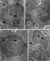
Electron micrographs illustrating morphological alterations in the basolateral amydaloid nucleus (ac) and in the ventral horns of the lumbar spinal cord (d) in 12-month-old GluR-B(N) mice. The neuron in a appears shrunken, and its cytoplasm has darkened and is filled with vacuolar structures. The arrows mark dilated cisternae of the endoplasmic reticulum, and the asterisks indicate swollen mitochondria. In b, a magnified view of the boxed area in a is shown. The dark pigment is lipofuscin, and the vacuoles can be identified as swollen mitochondria containing remnants of the mitochondrial cristae. A neuron from basolateral amygdaloid nucleus in c displays advanced morphological changes, although synaptic densities at its cytoplasmic membrane remain intact. Preservation of synaptic contacts and degeneration of postsynaptic structures is a hallmark neuropathological feature of excitotoxic neurodegeneration. Spinal motoneuron depicted in d displays pathological alterations consisting of darkening of the cytoplasm, formation of cytoplasmic vacuoles, which derive from the endoplasmic reticulum (arrows) or from the mitochondria, and shrinkage. (Scale bars: a and c, 1 μm; b, 200 μm; d, 5 μm.)
Fig. 4.
Fig. 4.
GluR-B expression and cobalt uptake in spinal cord of WT and GluR-B(N) transgenic mice. (a) mRNA in situ hybridization of lumbar spinal cord of GluR-B(N) and WT mice with a pan-probe recognizing native and transgenic GluR-B mRNA (total GluR-B mRNA) and with a probe specifically recognizing the GluR-B(N) transgene [GluR-B(N) mRNA]. (b) Quantification of positive grains corresponding to total GluR-B mRNA or GluR-B(N) mRNA in spinal motoneurons of WT and GluR-B(N) mice. Ventral horn neurons of GluR-B(N) mice show an increase in GluR-B mRNA-positive grains over WT mice. Furthermore, transgenic GluR-B(N) mice, but not WT mice, show specific expression of the GluR-B(N) transgene. (Scale bar: 100 μm.) (c) Immunostaining of the lumbar spinal cords of 4-month-old WT and age-matched GluR-B(N) transgenic mice with an anti-GluR-B antibody that recognizes both native GluR-B and GluR-B(N) transgene protein. (d) Boxed areas of ventral horns from c shown at a higher magnification. GluR-B(N) mice show significant increase in GluR-B immunoreactivity. (e) Labeling of functional AMPA receptors in the ventral horns of lumbar spinal cord of GluR-B(N) transgenic and WT mice by means of ligand-induced cobalt uptake. (Scale bar: 100 μm.) (f) Quantification of cobalt-positive cells in ventral horns from GluR-B(N) transgenic and WT mice. The number of neurons demonstrating functional AMPA receptors is significantly higher in the spinal ventral horn laminae VII and IX in GluR-B(N) transgenic vs. WT mice. (Scale bar: 100 μm.)

References

    1. Rowland, L. P. & Schneider, N. A. (2001) N. Engl. J. Med. 344, 1688–1700. - PubMed
    1. Bruijn, L. I., Miller, T. M. & Cleveland, D. W. (2004) Annu. Rev. Neurosci. 27, 723–749. - PubMed
    1. Couratier, P., Hugon, J., Sindou, P., Vallat, J. M. & Dumas, M. (1993) Lancet 341, 265–268. - PubMed
    1. Kawahara, Y., Ito, K., Sun, H., Aizawa, H., Kanazawa, I. & Kwak, S. (2004) Nature 427, 801. - PubMed
    1. Burnashev, N., Monyer, H., Seeburg, P. H. & Sakmann, B. (1992) Neuron 8, 189–198. - PubMed

MeSH terms