. 2005 Aug 1;106(3):841-51.
doi: 10.1182/blood-2004-05-2017. Epub 2005 Apr 12.

Preferential suppression of trisomy 8 compared with normal hematopoietic cell growth by autologous lymphocytes in patients with trisomy 8 myelodysplastic syndrome

Affiliations

Preferential suppression of trisomy 8 compared with normal hematopoietic cell growth by autologous lymphocytes in patients with trisomy 8 myelodysplastic syndrome

Elaine M Sloand et al. Blood. .

Abstract

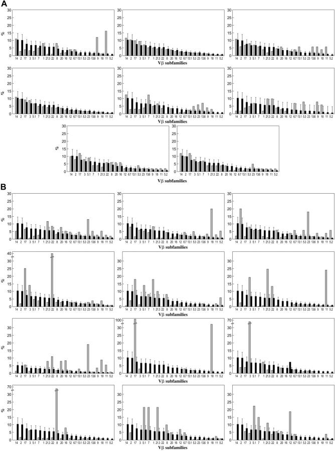
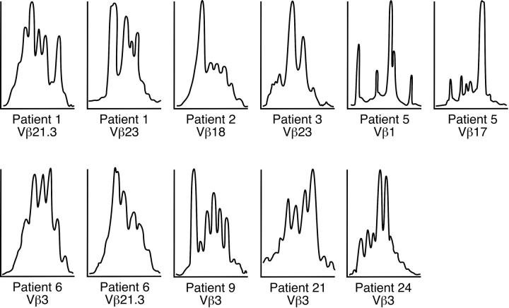
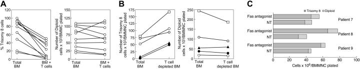
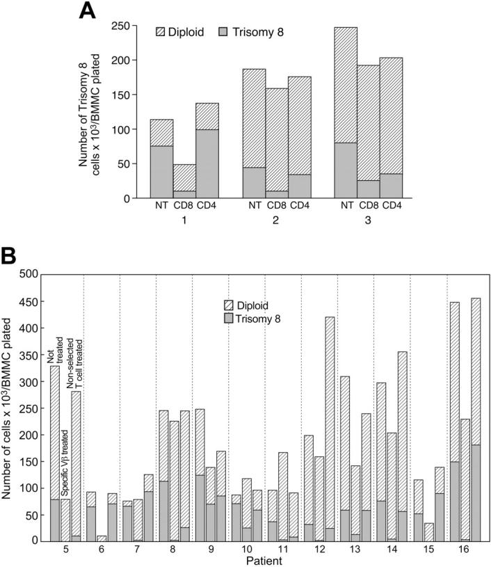
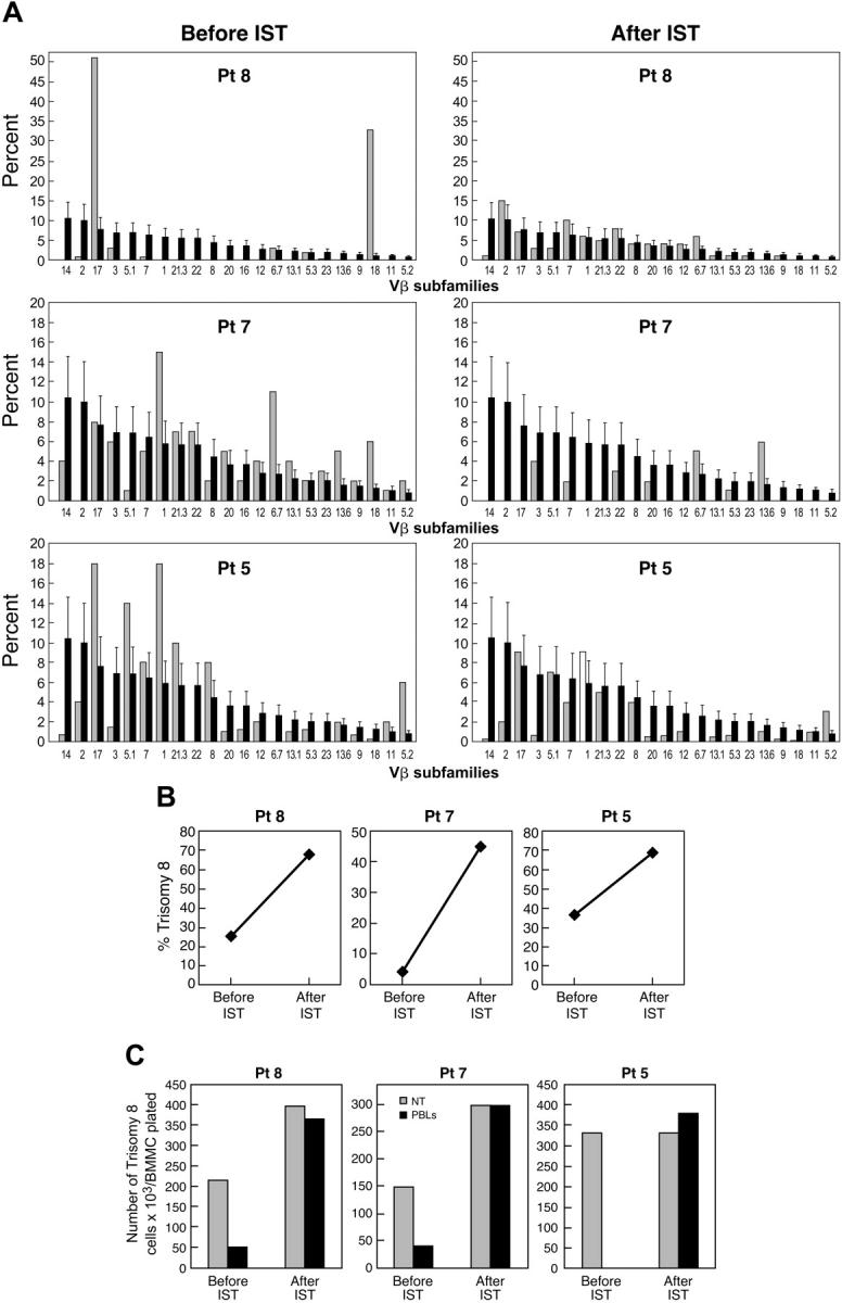
Clinical observations and experimental evidence link bone marrow failure in myelodysplastic syndrome (MDS) with a T cell-dominated autoimmune process. Immunosuppressive therapy is effective in improving cytopenias in selected patients. Trisomy 8 is a frequent cytogenetic abnormality in bone marrow cells in patients with MDS, and its presence has been associated anecdotally with good response to immunotherapy. We studied 34 patients with trisomy 8 in bone marrow cells, some of whom were undergoing treatment with antithymocyte globulin (ATG). All had significant CD8+ T-cell expansions of one or more T-cell receptor (TCR) Vbeta subfamilies, as measured by flow cytometry; expanded subfamilies showed CDR3 skewing by spectratyping. Sorted T cells of the expanded Vbeta subfamilies, but not of the remaining subfamilies, inhibited trisomy 8 cell growth in short-term hematopoietic culture. The negative effects of Vbeta-expanded T cells were inhibited by major histocompatibility complex (MHC) class 1 monoclonal antibody (mAb) and Fas antagonist and required direct cell-to-cell contact. Sixty-seven percent of patients who had de novo MDS with trisomy 8 as the sole karyotypic abnormality responded to ATG with durable reversal of cytopenias and restoration of transfusion independence, with stable increase in the proportion of trisomy 8 bone marrow cells and normalization of the T-cell repertoire. An increased number of T cells with apparent specificity for trisomy 8 cells is consistent with an autoimmune pathophysiology in trisomy 8 MDS.


Figures

Figure 1.
Figure 1.
TCR repertoire in trisomy 8 and monosomy 7. Thirty-three patients (light bars) with trisomy 8 (including 5 with complex cytogenetics), 11 patients with monosomy 7, and 19 age-matched healthy donors (dark bars) were studied. PBMC preparations were stained with CD28 FITC- and PE-conjugated mAbs directed against individual TCR-Vβ subfamilies and subjected to flow cytometry. Patients were compared with 19 age-matched controls. (A) Vβ subfamily distributions for a selection of patients with monosomy 7, only one of whom showed evidence of T-cell expansion. Values for each patient sample (light bars) are superimposed on mean of normal cohort (dark bars). (B) Examples of the Vβ subfamily distributions of patients with trisomy 8, all of whom showed expansion of Vβ subfamilies.
Figure 2.
Figure 2.
Spectratyping performed on trisomy 8 samples. Spectratyping demonstrated skewing of the CDR3 distribution in most expanded CD8+ Vβ subfamilies shown in Table 2.
Figure 3.
Figure 3.
Autologous lymphocytes decrease the proportion of BM trisomy B cells while Fas antagonist increases their growth. Fas agonist increases the proportion of trisomy 8 cells in short-term culture. BMMCs were obtained from 10 patients with trisomy 8, 4 patients with monosomy 7, and 4 healthy controls. Samples were divided into 2 aliquots, one of which was incubated with autologous PBLs for 4 hours, as described in “Patients, materials, and methods.” Samples were subsequently depleted of lymphocytes and placed in semisolid media with growth factors for short-term culture. Colonies were counted, and FISH was performed. The number of trisomy 8 cells and the percentage of trisomy 8 cells were decreased by lymphocyte cocultivation, with little effect seen in diploid cells (A). When one aliquot was depleted of T cells using CD3-specific magnetic beads before short-term culture and compared with the non–T cell–depleted sample, T-cell cultures consistently showed increased trisomy 8 progenitor–derived cell growth (B, left). T-cell depletion had little effect on the growth of cytogenetically normal cells (B, right). When samples of BM were plated in long-term culture with autologous lymphocytes, with and without Fas antagonist (ZB4 mAb), trisomy 8 cells increased compared to karyotypically normal cells (C).
Figure 4.
Figure 4.
Preferential inhibition of trisomy 8 cell colony growth related to suppression of growth by Vβ subfamily expanded CD8+ cells. Autologous column-purified CD4+ or CD8+ cells were incubated with 3 BMMC aliquots for 4 hours and placed in semisolid media for short-term culture in an additional 3 patients. Suppression of trisomy 8 progenitor–derived cell growth is seen for the BM cells previously incubated with CD8+ cells only (A). Similar experiments performed with Vβ-selected CD8+ cells (B) from an additional 7 patients showed preferential inhibition by selected, but not unselected, cells of trisomy 8 but not karyotypically normal cells. Erythroid and myeloid cells were affected.
Figure 5.
Figure 5.
Clinical development or disappearance of trisomy 8 associated with presence or absence of Vβ expansion. (A) Flow cytometric examination of Vβ subfamilies from patients 5, 7, and 8 before and after IST. Values for each patient (light bars) are superimposed on mean values of normal cohort (dark bars). (B) FISH data showing percentage of trisomy 8 before and after IST. (C) Effect of lymphocytes on trisomy 8 cell growth before and after IST. Paired lymphocytes and BMMCs obtained before and after IST were incubated in close contact with effector cells (with effector-target ratios of 2:1) for 2 hours before placement in short-term culture for 14 days. When FISH was performed, the number of trisomy 8 cells was decreased by cocultivation with the lymphocytes obtained before IST only.

Comment in

References

    1. Bennett JM. World Health Organization classification of the acute leukemias and myelodysplastic syndrome. Int J Hematol. 2000;72: 131-133. - PubMed
    1. Deeg HJ, Beckham C, Loken MR, et al. Negative regulators of hemopoiesis and stroma function in patients with myelodysplastic syndrome. Leuk Lymphoma. 2000;37: 405-414. - PubMed
    1. Parker JE, Fishlock KL, Mijovic A, et al.'Low-risk' myelodysplastic syndrome is associated with excessive apoptosis and an increased ratio of proversus anti-apoptotic bcl-2-related proteins. Br J Haematol. 1998;103: 1075-1082. - PubMed
    1. Parker JE, Mufti GJ. Ineffective haemopoiesis and apoptosis in myelodysplastic syndromes. Br J Haematol. 1998;101: 220-230. - PubMed
    1. Parker JE, Mufti GJ. Excessive apoptosis in low risk myelodysplastic syndromes (MDS). Leuk Lymphoma. 2000;40: 1-24. - PubMed

MeSH terms

Substances