Comparative Study
. 2005 May;79(9):5288-95.
doi: 10.1128/JVI.79.9.5288-5295.2005.

Identification of novel subgenomic RNAs and noncanonical transcription initiation signals of severe acute respiratory syndrome coronavirus

Affiliations
Comparative Study

Identification of novel subgenomic RNAs and noncanonical transcription initiation signals of severe acute respiratory syndrome coronavirus

Snawar Hussain et al. J Virol. 2005 May.

Abstract

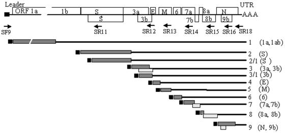
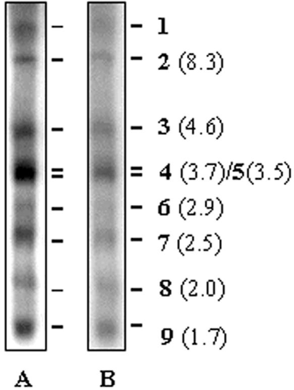
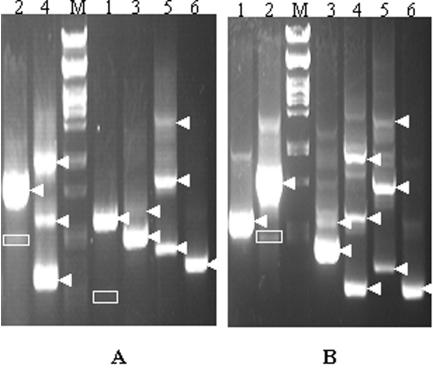
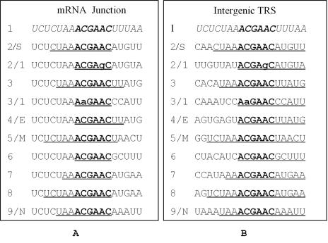
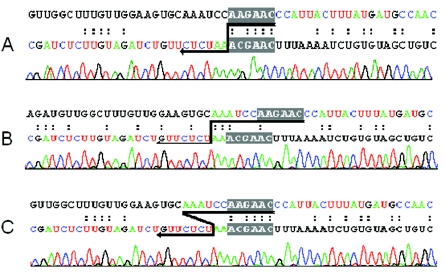
The expression of the genomic information of severe acute respiratory syndrome coronavirus (SARS CoV) involves synthesis of a nested set of subgenomic RNAs (sgRNAs) by discontinuous transcription. In SARS CoV-infected cells, 10 sgRNAs, including 2 novel ones, were identified, which were predicted to be functional in the expression of 12 open reading frames located in the 3' one-third of the genome. Surprisingly, one new sgRNA could lead to production of a truncated spike protein. Sequence analysis of the leader-body fusion sites of each sgRNA showed that the junction sequences and the corresponding transcription-regulatory sequence (TRS) are unique for each species of sgRNA and are consistent after virus passages. For the two novel sgRNAs, each used a variant of the TRS that has one nucleotide mismatch in the conserved hexanucleotide core (ACGAAC) in the TRS. Coexistence of both plus and minus strands of SARS CoV sgRNAs and evidence for derivation of the sgRNA core sequence from the body core sequence favor the model of discontinuous transcription during minus-strand synthesis. Moreover, one rare species of sgRNA has the junction sequence AAA, indicating that its transcription could result from a noncanonical transcription signal. Taken together, these results provide more insight into the molecular mechanisms of genome expression and subgenomic transcription of SARS CoV.


Figures

FIG. 1.
FIG. 1.
Schematic representation of the genomic and subgenomic organizations of SARS CoV. The genome organization is based on the sequence of the SARS WHU isolate. In the upper panel, the genomic structure is shown. Known and potential ORFs are indicated by open boxes and are not to scale. The leader region is represented by a small solid box, and the poly(A) tail is represented by AAA. ORF 8a of isolate WHU contains a 2-nucleotide deletion and thus gives rise to a small ORF of 24 amino acids. Positions of forward (SF8 and -9) and reverse (SR11 to -18) primers used for cDNA synthesis and PCR amplification of different subgenomic RNAs are indicated by arrows under the genome. The bottom panel illustrates the 3′-coterminal nested set of mRNAs detected in this study. The small black boxes at the 5′ ends of the genomic and subgenomic RNAs represent the common leader sequence. The first (grey boxes) and second (open boxes) ORFs that are located at the 5′-proximal end and may be expressed from the mRNAs are shown.
FIG. 2.
FIG. 2.
Northern blot analysis of SARS CoV subgenomic mRNAs Twenty micrograms of total cellular RNA from SARS CoV-infected Vero E6 cells was separated by electrophoresis through a 1.2% denaturing agarose gel containing 2.2 M formaldehyde. The resolved RNA was transferred to a nylon membrane, and 32P-labeled antisense and sense probes containing 305 nucleotides (positions 29421 to 29725) from the 3′ end of the SARS CoV genome were used to detect subgenomic mRNAs and minus-strand subgenomic RNAs, respectively. The mRNA designations and their approximate sizes (in parentheses) are indicated on the right. (A) Subgenomic mRNAs detected by the negative probe; (B) minus-strand subgenomic RNAs detected by the positive probe.
FIG. 3.
FIG. 3.
RT-PCR analysis of SARS CoV subgenomic RNAs. One microgram of total cellular RNA from SARS CoV-infected Vero E6 cells was reverse transcribed and amplified by PCR with different combinations of forward (SF9) and reverse (SP11 to SP16 [lanes 1 to 6, respectively]) primers. The bands representing the specific SARS CoV sequences are indicated by arrowheads. The bands which revealed two novel subgenomic RNAs are boxed. Lanes 1, mRNA 2 (arrowhead) and mRNA 2-1 (boxed faint band in panel A); lanes 2, mRNA 3 (major band) and mRNA 3-1 (boxed); lanes 3, mRNA 5 (lower major band) and 4 (upper minor band); lanes 4, mRNA 7 (lower band), mRNA 6 (middle band), and mRNA 5 (upper band); lanes 5, mRNA 8 (lower band), mRNA 7 (middle band), and mRNA 6 (upper band); lane 6, mRNA 9. (A). The cDNA used for PCR was made with oligo(dT) or SR18 primer, and thus the sequence of corresponding plus-strand RNA was amplified. (B). The cDNA used for PCR was made with primer SF8, which is complementary to the antileader sequence, and therefore the sequence of corresponding minus-strand RNA was amplified.
FIG. 4.
FIG. 4.
Leader-body fusion sites of subgenomic mRNAs and their corresponding intergenic sequences. The 5′ genomic leader TRS is in italic. The hexanucleotide core sequence of the TRS is indicated in boldface, and the mismatched nucleotides with the leader core sequence (ACGAAC) are in lowercase. (A) Leader-body junction sites of subgenomic mRNAs in comparison with the genomic leader TRS. The junction sequences in subgenomic RNAs are underlined. (B) The TRS in the intergenic regions. The body sequences that are fused with the 5′ leader are underlined.
FIG. 5.
FIG. 5.
Junction sequences of SARS CoV mRNA 3-1 and models for template switch. The upper strand in the alignments represents the intergenic region of mRNA 3-1, and the lower strand is the genomic leader sequence. Dots indicate identity between the sequences. The conserved hexanucleotide core sequence is shaded, and the possible site for the template switch is indicated by arrow. The nucleotides in color are derived directly from the sequence profiles. (A) Leader-body fusion site of mRNA 3-1; (B and C) junction sequence and models of template switch of a rare variant of mRNA 3-1.

References

    1. An, S., A. Maeda, and S. Makino. 1998. Coronavirus transcription early in infection. J. Virol. 72:8517-8524. - PMC - PubMed
    1. Baric, R. S., and B. Yount. 2000. Subgenomic negative-strand RNA function during mouse hepatitis virus infection. J. Virol. 74:4039-4046. - PMC - PubMed
    1. Callebaut, P., I. Correa, M. Pensaert, G. Jimenez, and L. Enjuanes. 1988. Antigenic differentiation between transmissible gastroenteritis virus of swine and a related porcine respiratory coronavirus. J. Gen. Virol. 69:1725-1730. - PubMed
    1. Chinese SARS Molecular Epidemiology Consortium. 2004. Molecular evolution of the SARS coronavirus during the course of the SARS epidemic in China. Science 303:1666-1669. - PubMed
    1. De Vries, A. F., M. C. Horzinek, P. J. M. Rottier, and R. J. de Groot. 1997. The genome organization of the Nidovirales: similarities and differences between arteri-, toro-, and coronaviruses. Semin. Virol. 8:33-47. - PMC - PubMed

Publication types

LinkOut - more resources