. 2005 May;79(9):5326-36.
doi: 10.1128/JVI.79.9.5326-5336.2005.

RhoA signaling is required for respiratory syncytial virus-induced syncytium formation and filamentous virion morphology

Affiliations

RhoA signaling is required for respiratory syncytial virus-induced syncytium formation and filamentous virion morphology

Tara L Gower et al. J Virol. 2005 May.

Abstract

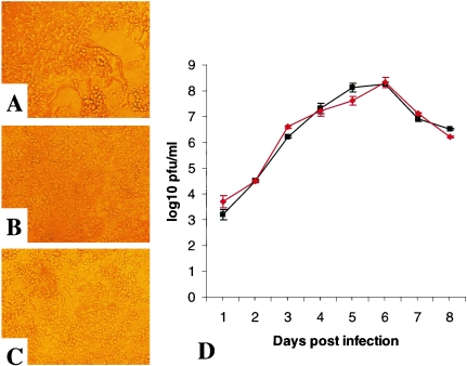
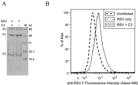
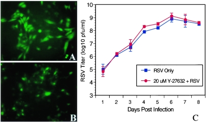
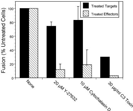
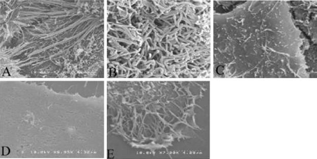
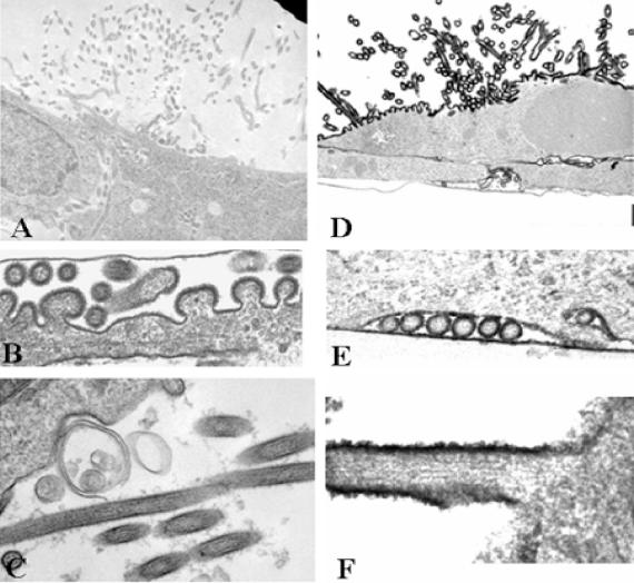
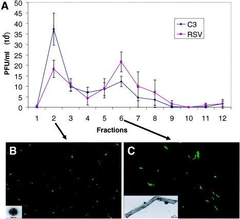
Respiratory syncytial virus (RSV) is an important human pathogen that can cause severe and life-threatening respiratory infections in infants, the elderly, and immunocompromised adults. RSV infection of HEp-2 cells induces the activation of RhoA, a small GTPase. We therefore asked whether RhoA signaling is important for RSV replication or syncytium formation. The treatment of HEp-2 cells with Clostridium botulinum C3, an enzyme that ADP-ribosylates and specifically inactivates RhoA, inhibited RSV-induced syncytium formation and cell-to-cell fusion, although similar levels of PFU were released into the medium and viral protein expression levels were equivalent. Treatment with another inhibitor of RhoA signaling, the Rho kinase inhibitor Y-27632, yielded similar results. Scanning electron microscopy of C3-treated infected cells showed reduced numbers of single blunted filaments, in contrast to the large clumps of long filaments in untreated infected cells. These data suggest that RhoA signaling is associated with filamentous virus morphology, cell-to-cell fusion, and syncytium formation but is dispensable for the efficient infection and production of infectious virus in vitro. Next, we developed a semiquantitative method to measure spherical and filamentous virus particles by using sucrose gradient velocity sedimentation. Fluorescence and transmission electron microscopy confirmed the separation of spherical and filamentous forms of infectious virus into two identifiable peaks. The C3 treatment of RSV-infected cells resulted in a shift to relatively more spherical virions than those from untreated cells. These data suggest that viral filamentous protuberances characteristic of RSV infection are associated with RhoA signaling, are important for filamentous virion morphology, and may play a role in initiating cell-to-cell fusion.


Figures

FIG. 1.
FIG. 1.
C3 prevents RSV-induced syncytium formation in HEp-2 cells. (A) RSV-infected cells showed extensive syncytium formation in HEp-2 cells at 3 days postinfection. (B) Treatment with 30 μg of C3/ml blocked RSV-induced syncytium formation. (C) Uninfected control HEp-2 cells treated with 30 μg of C3/ml showed that C3 has no visible effect on cell morphology or viability. (D) HEp-2 cell monolayers grown in 96-well plates were either treated with 30 μg of C3/ml for 24 h or mock treated and then were infected with RSV at an MOI of 0.1. Virus titers were measured for eight consecutive days after RSV infection by harvesting the entire contents of each well and performing plaque assays in triplicate. RSV growth curves were created for untreated cells (squares) and cells treated with C3 (diamonds). The data shown are representative of three separate experiments. Error bars represent standard deviations.
FIG. 2.
FIG. 2.
Level of F expression in C3-treated or untreated infected cells. (A) HEp-2 cells were either left untreated or pretreated with 30 μg of C3/ml 24 h prior to RSV infection. At 72 h postinfection, the cells were harvested and a 1-ml aliquot was centrifuged at 14,000 rpm for 10 min at 4°C. The pellet was resuspended in mammalian protein extraction reagent. Equal amounts of proteins from C3-treated or untreated RSV-infected cells were resolved by sodium dodecyl sulfate-10% polyacrylamide gel electrophoresis and transferred to polyvinylidene difluoride membranes. The F protein was detected with an anti-F monoclonal antibody followed by an HRP-conjugated anti-mouse antibody. Proteins were visualized by ECL. +, C3 treatment; −, no treatment. (B) RSV F expression. C3-treated cells infected with RSV for 24 h were evaluated by flow cytometry.
FIG. 3.
FIG. 3.
Effect of Y-27632 on RSV replication. rgRSV-infected cells can be visualized by immunofluorescence microscopy. (A) At 24 h postinfection, rgRSV-infected cultures had many green infected single cells. (B) Pretreatment with 20 μM Y-27632 for 24 h did not affect the single-cell infection by rgRSV. (C) HEp-2 cell monolayers grown in 96-well plates were either treated with 20 μM Y-27632 or left untreated and infected with RSV at an MOI of 0.1. Virus titers were measured for eight consecutive days after RSV infection by harvesting the entire contents of each well and performing plaque assays in triplicate. RSV growth curves were created for untreated cells (squares) and cells treated with 20 μM Y-27632 (circles). The data shown are representative of three separate experiments. Error bars represent standard deviations.
FIG. 4.
FIG. 4.
Role of RhoA-induced signaling in RSV-induced cell-to-cell fusion. Fusion was measured by combining effector cells (HEp-2 cells infected with a vaccinia virus expressing T7 polymerase and transfected with plasmids encoding RSV F, G, and SH) with target cells (HEp-2 cells infected with a vaccinia virus expressing lacZ under the control of a T7 promoter). The cells were fixed 4 h after mixing and stained with X-Gal for the visualization of blue fused cells. Prior to mixing, either target cells (black bars) or effector cells (hatched bars) were treated with C3, Y-27632, or cytochalasin D for 16 h beginning 4 h after vaccinia virus infection or transfection and then washed.
FIG. 5.
FIG. 5.
Inactivating RhoA causes blunted viral filaments. Viral filaments were visualized by scanning electron microscopy. Cells infected with RSV for 24 h (A) or 48 h (B) had large clumps of filaments protruding from the cells. HEp-2 cells treated with 30 μg of C3/ml and infected with RSV (C) had reduced quantities of single blunted filaments at 24 h, while uninfected HEp-2 cells (D) had no filamentous structures. The filaments in RSV-infected cells resembled RhoA-induced microvilli in uninfected cells treated with 20 μM LPA for 30 min (E). Magnification, ×6,300.
FIG. 6.
FIG. 6.
Ultrastructural localization of RSV F protein on viral particles and filaments. Vero cells infected with the Long strain of RSV were fixed at 48 h postinfection and processed for immunoelectron microscopic localization of the F protein. The cells in panels A to C were control immunostained cultures grown in the absence of the F-specific monoclonal antibody SB-209763, while the cells in panels D to F were grown in the presence of 10 μg of SB-209763/ml, which was localized with HRP-conjugated donkey anti-human IgG. Viral particles (B and E) and filaments (C and F) had viral glycoprotein spikes on their surfaces (B and C) that stained positively with the anti-F antibody (E and F). Nucleocapsid structures are evident within both viral particles and filaments. Bar = 1 μm (A and D) or 100 nm (B, C, E, and F).
FIG. 7.
FIG. 7.
Sucrose gradient analysis of virus particles. Velocity sedimentation was performed on RSV grown for 72 h and treated with 30 μg of C3/ml or left untreated. In each of three independent experiments, C3-treated cells produced more infectious RSV in the first peak and less in the second peak of the gradient. (A) Representative graph of the PFU in each fraction. Evaluating the results of three separate experiments, we found the ratio of untreated to C3-treated PFU for peak 1 to be 1.80 ± 0.18 and that for peak 2 to be 0.58 ± 0.001 (two-tailed t test; P < 0.01). Using this technique, we examined the morphology of virions from peaks 1 and 2 by fluorescence microscopy using anti-F monoclonal antibody, followed by Alexafluor 488 anti-mouse IgG. The images shown represent untreated RSV, but the morphologies of the two peaks for C3-treated RSV were similar. The first peak contained predominantly spherical or pleomorphic virus particles (B), and the second peak contained primarily filamentous particles (C). The identities of the morphologically distinct virus particles were confirmed by electron microscopy (insets). These data indicate that a C3 treatment shifts the virion morphology to a more spherical and less filamentous shape.

References

    1. Anderson, H. A., Y. Chen, and L. C. Norkin. 1996. Bound simian virus 40 translocates to caveolin-enriched membrane domains, and its entry is inhibited by drugs that selectively disrupt caveolae. Mol. Biol. Cell 7:1825-1834. - PMC - PubMed
    1. Bachi, T. 1988. Direct observation of the budding and fusion of an enveloped virus by video microscopy of viable cells. J. Cell Biol. 107:1689-1695. - PMC - PubMed
    1. Bachi, T., and C. Howe. 1973. Morphogenesis and ultrastructure of respiratory syncytial virus. J. Virol. 12:1173-1180. - PMC - PubMed
    1. Bergelson, J. M., and R. W. Finberg. 1993. Integrins as receptors for virus attachment and cell entry. Trends Microbiol. 1:287-288. - PubMed
    1. Brown, G., H. W. Rixon, and R. J. Sugrue. 2002. Respiratory syncytial virus assembly occurs in GM1-rich regions of the host-cell membrane and alters the cellular distribution of tyrosine-phosphorylated caveolin-1. J. Gen. Virol. 83:1841-1850. - PubMed

Publication types

MeSH terms

LinkOut - more resources