. 2005 May;79(9):5386-99.
doi: 10.1128/JVI.79.9.5386-5399.2005.

Covert human immunodeficiency virus replication in dendritic cells and in DC-SIGN-expressing cells promotes long-term transmission to lymphocytes

Affiliations

Covert human immunodeficiency virus replication in dendritic cells and in DC-SIGN-expressing cells promotes long-term transmission to lymphocytes

Cinzia Nobile et al. J Virol. 2005 May.

Abstract

HIV-1 virions are efficiently captured by monocyte-derived immature dendritic cells (iDCs), as well as by cell lines expressing the lectin DC-SIGN. Viral infectivity can be retained for several days, and even enhanced, before transmission to CD4+ lymphocytes. The role of DC-SIGN in viral retention and enhancement of infection is not fully understood and varies according to the cell line expressing the lectin. We studied here the mechanisms underlying this process. We focused our study on X4-tropic human immunodeficiency virus (HIV) strains, since they were widely believed not to replicate in iDCs. However, we first show that X4 HIV replicates covertly and slowly in iDCs. This is also the case in Raji-DC-SIGN cells, which are classically used to study HIV transmission. We used either single-cycle or replicative HIV and measured viral RT and replication to further demonstrate that transfer of incoming virions from iDCs or DC-SIGN+ cells occurs only on the short-term (i.e., a few hours after viral exposure). There is no long-term storage of original HIV particles in these cells. A few days after viral exposure, replicative viruses, and not single-cycle virions, are transmitted to CD4+ cells. The cell-type-dependent activity of DC-SIGN reflects the ability of HIV to replicate covertly in some cells, and not in others.


Figures

FIG. 1.
FIG. 1.
HIV replication and proviral DNA synthesis in monocyte-derived iDCs. (A) HIV replication. iDCs (106 cells) were exposed to the indicated viruses (50 ng of p24), with or without AZT (10 μM). After overnight incubation, cells were washed to remove unbound virus. Viral replication was monitored by measuring p24 production in culture supernatants. One of six independent experiments is shown. (B and C) HIV proviral DNA synthesis. iDCs were exposed to the indicated viruses (50 ng of p24/106 cells) for 3 h and extensively washed. Cell aliquots were then immediately collected or incubated at 37°C for various periods of time. Quantification of early (RU5 DNA) (B) and late (Pol DNA) (C) viral products was performed by real-time PCR. Data are means ± standard deviation of triplicates and are representative of at least three independent experiments, performed with DCs from different donors.
FIG. 2.
FIG. 2.
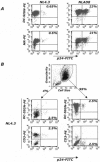
Intracellular p24 expression by HIV-infected iDCs. (A) DCs from one donor were exposed to the indicated viruses (50 ng of p24/106 cells) for 2 h at 37°C. Five days later, fluorescence-activated cell sorter analysis was performed to monitor HIV infection (p24-FITC staining, x axis) and the surface proteins DC-SIGN and MR (phycoerythrin staining, y axis). (B) iDCs from another donor were exposed to the X4 strain NL4.3 (50 ng of p24/106 cells). Five days later, cells were stained for p24, DC-SIGN, MR, or CD3. Similar results were obtained with cells from four different donors.
FIG. 3.
FIG. 3.
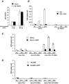
HIV RT in iDCs requires viral fusion. (A) Capture of WT (NL4.3) and fusion-defective (NL F522Y) HIV by iDCs. Cells were exposed to the indicated viruses (6 or 30 ng of p24/106 cells) for 2 h at 37°C and washed extensively, and cell-associated p24 was quantified by ELISA. (B) RT in iDCs. Cells were exposed to the indicated viruses as described in the legend to Fig. 1. Quantification of RU5 viral DNA was performed by real time PCR. (C-D) RT in HeLa cells. Cells were transiently transfected with CD4 and/or DC-SIGN and were exposed to NL4.3 (C) or NLAD8 (D). RU5 viral products were quantified by real-time PCR as described in the legend to Fig. 1. Data are means ± standard deviation of triplicates and are representative of at least three independent experiments.
FIG. 4.
FIG. 4.
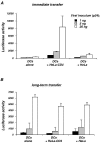
Capture and transmission of incoming HIV by iDCs. iDCs (106 cells) were exposed to the indicated doses of single-cycle HIV-Luc (pseudotyped with X4 envelope glycoproteins) and extensively washed. Cells were then either cultured alone or with target HeLa or HeLa-CD4 cells. Cocultures were performed either immediately (immediate transfer) (A) or 48 h after viral exposure (long-term transfer) (B). Cell lysates were obtained after an additional 48-h culture period and analyzed for luciferase activity (in relative light units). Data represent the means ± standard deviation of three separate wells of infected cells and are representative of at least three independent experiments.
FIG. 5.
FIG. 5.
Transmission of incoming HIV from iDCs to T cells. iDCs were exposed to single-cycle HIV-Luc (pseudotyped with X4 envelope glycoproteins) (100 ng of p24/1.5 × 106 cells) and extensively washed. Cells were then either cultured alone or with target Jurkat (left) or MT4 (right) T cells. Cocultures were initiated at the indicated time points. Cell lysates were obtained after an additional 48-h culture period and analyzed for luciferase activity (in relative light units). Data represent means ± standard deviation of three separate wells of infected cells and are representative of three independent experiments.
FIG. 6.
FIG. 6.
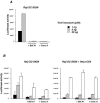
Capture and transmission of incoming HIV by DC-SIGN-expressing B cell lines. (A) Transmission of single-cycle HIV-Luc. Raji DC-SIGN (top) and C1RA2 DC-SIGN (bottom) cells were exposed to the indicated doses of HIV-Luc. Cells were then either cultured alone or with target HeLa or HeLa-CD4 cells. Cocultures were performed either immediately (immediate transfer) or 48 h after viral exposure (long-term transfer). Cell lysates were obtained after an additional 48-h culture period and analyzed for luciferase activity. Data represent means ± standard deviation of triplicates and are representative of at least three independent experiments. (B) Transmission of replicative HIV. Raji DC-SIGN (top) and C1RA2 DC-SIGN (bottom) were exposed to the indicated doses of NL4.3, washed, and cocultivated with P4 cells (HeLa-CD4 LTR-LacZ reporter cells) either immediately or 48 h after viral exposure. HIV transmission to P4 cells was assessed by measuring β-galactosidase activity in cell extracts after a 2-day coculture. Data (measured by optical density) are means ± standard deviation of triplicates and are representative of three independent experiments.
FIG. 7.
FIG. 7.
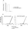
HIV proviral DNA synthesis in Raji DC-SIGN cells. Raji and Raji DC-SIGN cells were exposed to NL4.3 (50 ng of p24/106 cells) as described in the legend to Fig. 1. Quantification of RU5 viral DNA was performed by real-time PCR. Data are means ± standard deviation of triplicates and are representative of three independent experiments.
FIG. 8.
FIG. 8.
pH-independent HIV transmission by Raji DC-SIGN cells. (A) Infection by HIV(VSV) pseudotypes is inhibited by bafilomycin A1 and concanamycin A. Raji DC-SIGN cells were pretreated or not pretreated with bafilomycin A1 (Baf A1, 250 nM) or with concanamycin A (Conc A, 10 nM) for 1 h and then pulsed with the indicated doses of HIV-Luc pseudotyped with VSV-G [HIV-Luc(VSV)] for 2 h with or without the drugs. Luciferase activity in cell lysates was measured 2 days later. (B) Effect of bafilomycin A1 and concanamycin A on HIV infection and transmission by Raji DC-SIGN cells. Raji DC-SIGN cells were pretreated or not pretreated with bafilomycin A1 or concanamycin A and then pulsed with the indicated doses of HIV-Luc as described for panel A). Cells were then grown alone (left) or cocultivated with target HeLa-CD4 cells (right). Luciferase activity was measured 2 days later in cell lysates. One out of three independent experiments is shown.
FIG. 9.
FIG. 9.
pH-independent HIV transmission by iDCs. (A) Infection of iDCs by HIV(VSV) pseudotypes is inhibited by bafilomycin A1. iDCs were pretreated or not pretreated with bafilomycin A1 (Baf A1, 250 nM) for 1 h, and then pulsed with HIV-Luc(VSV) (20 ng of p24/106 cells). Luciferase activity was measured 2 days later in cell lysates. (B) Bafilomycin A1 does not affect HIV transmission from iDCs to Jurkat cells. iDCs (5 × 10⁵ cells) were pretreated or not pretreated for 1 h with bafilomycin A1 or mock treated, exposed to 0.1 (left) or 0.01 (right) ng of NL4./ml for 2 h, washed, and cocultivated with Jurkat cells (5 × 10⁵ cells). As a control, Jurkat cells were exposed to the same virus inputs and cultured alone. Viral replication was monitored by measuring p24 production in culture supernatants. One of three independent experiments is shown.
FIG. 10.
FIG. 10.
Replication of HIV strains carrying primary X4 and R5 envelope glycoproteins in iDCs. The primary envelope genes were obtained by RT-PCR amplification from a plasma sample issued from an HIV-infected patient and cloned in an env-deleted NL4.3 HIV isolate. Two X4 (T28-X4-1 and T28-X4-2) and one R5 (T28-R5-1) isolates were analyzed. iDCs were exposed to the indicated viruses (50 or 5 ng of p24/106 cells). After overnight incubation, cells were washed to remove unbound virus. Virus replication was monitored by measuring p24 production in culture supernatants. One of three independent experiments is shown.

References

    1. Aiken, C. 1997. Pseudotyping human immunodeficiency virus type 1 (HIV-1) by the glycoprotein of VSV targets HIV-1 entry to an endocytic pathway and suppresses both the requirement for Nef and the sensitivity to cyclosporin A. J. Virol. 71:5871-5877. - PMC - PubMed
    1. Arrighi, J. F., M. Pion, M. Wiznerowicz, T. B. Geijtenbeek, E. Garcia, S. Abraham, F. Leuba, V. Dutoit, O. Ducrey-Rundquist, Y. van Kooyk, D. Trono, and V. Piguet. 2004. Lentivirus-mediated RNA interference of DC-SIGN expression inhibits human immunodeficiency virus transmission from dendritic cells to T cells. J. Virol. 78:10848-10855. - PMC - PubMed
    1. Ayehunie, S., E. A. Garcia-Zepeda, J. A. Hoxie, R. Horuk, T. S. Kupper, A. D. Luster, and R. M. Ruprecht. 1997. Human immunodeficiency virus-1 entry into purified blood dendritic cells through CC and CXC chemokine coreceptors. Blood 90:1379-1386. - PubMed
    1. Bakri, Y., C. Schiffer, V. Zennou, P. Charneau, E. Kahn, A. Benjouad, J. C. Gluckman, and B. Canque. 2001. The maturation of dendritic cells results in postintegration inhibition of HIV-1 replication. J. Immunol. 166:3780-3788. - PubMed
    1. Baribaud, F., R. W. Doms, and S. Pohlmann. 2002. The role of DC-SIGN and DC-SIGNR in HIV and Ebola virus infection: can potential therapeutics block virus transmission and dissemination? Expert Opin. Ther. Targets 6:423-431. - PubMed

Publication types

MeSH terms

Substances