. 2005 May;17(5):1467-81.
doi: 10.1105/tpc.104.030155. Epub 2005 Apr 13.

Disruptions of the Arabidopsis Enoyl-CoA reductase gene reveal an essential role for very-long-chain fatty acid synthesis in cell expansion during plant morphogenesis

Affiliations

Disruptions of the Arabidopsis Enoyl-CoA reductase gene reveal an essential role for very-long-chain fatty acid synthesis in cell expansion during plant morphogenesis

Huanquan Zheng et al. Plant Cell. 2005 May.

Abstract

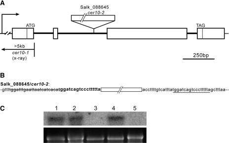
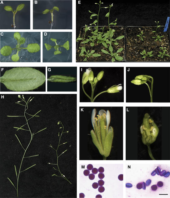
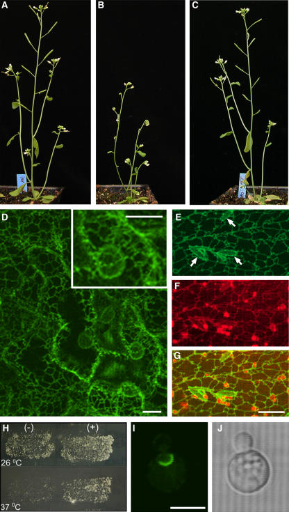
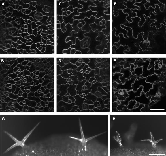
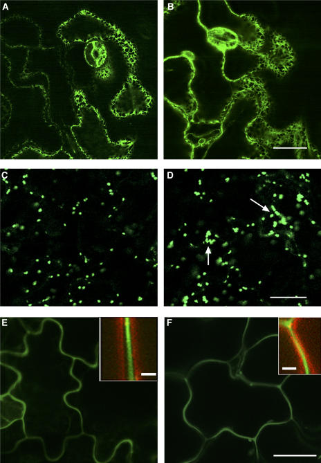
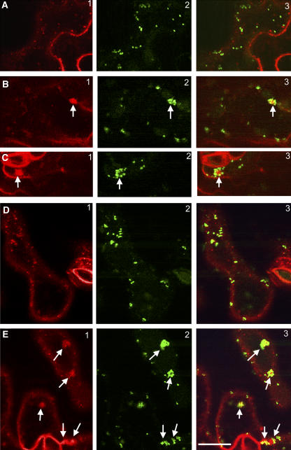
In the absence of cell migration, plant architecture is largely determined by the direction and extent of cell expansion during development. In this report, we show that very-long-chain fatty acid (VLCFA) synthesis plays an essential role in cell expansion. The Arabidopsis thaliana eceriferum10 (cer10) mutants exhibit severe morphological abnormalities and reduced size of aerial organs. These mutants are disrupted in the At3g55360 gene, previously identified as a gene coding for enoyl-CoA reductase (ECR), an enzyme required for VLCFA synthesis. The absence of ECR activity results in a reduction of cuticular wax load and affects VLCFA composition of seed triacylglycerols and sphingolipids, demonstrating in planta that ECR is involved in all VLCFA elongation reactions in Arabidopsis. Epidermal and seed-specific silencing of ECR activity resulted in a reduction of cuticular wax load and the VLCFA content of seed triacylglycerols, respectively, with no effects on plant morphogenesis, suggesting that the developmental phenotypes arise from abnormal sphingolipid composition. Cellular analysis revealed aberrant endocytic membrane traffic and defective cell expansion underlying the morphological defects of cer10 mutants.


Figures

Figure 1.
Figure 1.
Identification of cer10-1 and cer10-2 Mutations in the ECR Gene. (A) The ECR gene structure and mutation sites of cer10 alleles. White boxes represent exons. cer10-1 has at least a 5-kb deletion and/or rearrangement located in the promoter region, the 5′-untranslated region, and at least 90 bp downstream of the translation start of the gene, whereas cer10-2 (Salk_088645) carries a T-DNA insertion in the second intron of the gene. (B) The genomic DNA sequence flanking the T-DNA insertion in cer10-2. White box represents T-DNA insertion site. A small deletion of 22 bp (strikethrough) and a small duplication (bold) of an 18-bp genomic DNA fragment (underline) flanking the T-DNA insertion in cer10-2 are detected. (C) RNA gel blot analysis of steady state ECR mRNA in wild-type Col-0 (lane 1), Salk_138092 (lane 2), cer10-2 (lane 3), wild-type Ler (lane 4), and cer10-1 (lane 5). The bottom panel shows ethidium bromide–stained 18S rRNAs.
Figure 2.
Figure 2.
Abnormal Organ Morphogenesis in the cer10 Mutant. (A) and (B) Four-day old seedlings of wild-type Col-0 (A) and cer10-2 (B). (C) and (D) Twelve-day-old seedlings of wild-type Col-0 (C) and cer10-2 (D). (E) Twenty-two-day-old wild-type Col-0 (left) and cer10-2 (right) plants. (F) and (G) Expanded adult leaves of wild-type Col-0 (F) and cer10-2 (G). (H) Stems of adult wild-type Col-0 (left) and cer10-2 (right) plants. (I) and (J) Flowers of wild-type Col-0 (I) and cer10-2 (J). (K) and (L) Mechanically opened 11 to 13 stage flowers of wild-type Col-0 (K) and cer10-2 (L). (M) and (N) Pollen grains of wild-type Col-0 (M) and cer10-2 (N) labeled with Alexander's stain. Bar = 10 μm for (M) and (N).
Figure 3.
Figure 3.
Complementation of the cer10 Mutant and Subcellular Localization of the ECR. (A) to (C) Four-week-old wild-type Ler (A), cer10-1 (B), and cer10-1:GFP-ECR plants (C). (D) Projection of 12 × 1-μm optical sections of GFP fluorescence through GFP-ECR expressing cer10-1 leaf epidermal cells. Note the similar fluorescence intensity between cortical ER network and nuclear envelopes. Bars = 5 μm. (E) to (G) Colocalization of GFP-ECR labeled network (E) and the ER network stained by hexyl rhodamine B (F). (G) was obtained by merging images (E) and (F). The arrows in (E) identify the fusiform bodies of the ER outlined in negative contrast by GFP-ECR. Bar = 10 μm for (E) to (G). (H) The yeast tsc13-1elo2Δ cells transformed with the plasmid p426-ADH without GFP-ECR growing at 26°C (top left) and at 37°C (bottom left) and the tsc13-1elo2Δ cells transformed with the plasmid p426-ADH:GFP-ECR growing at 26°C (top right) and 37°C (bottom right). (I) and (J) Confocal GFP fluorescence (I) and bright-field (J) images of a yeast tsc13-1elo2Δ cell expressing GFP-ECR. Bar = 5 μm.
Figure 4.
Figure 4.
Defective Cell Expansion in the cer10 Mutants. (A) to (F) Cell shape was defined by FM4-64 staining, and all images were projections of 10 × 1-μm optical sections of FM4-64 fluorescence through leaf epidermal cells. Bar = 25 μm. (A) and (B) Newly formed epidermal cells of a wild-type Ler leaf (A) and a cer10-1 mutant leaf (B). (C) and (D) Expanding epidermal cells of a wild-type Ler leaf (C) and a cer10-1 mutant leaf (D). (E) and (F) Epidermal cells in a fully expanded wild-type Ler leaf (E) and a cer10-1 mutant leaf (F). (G) Stereomicrograph of wild-type Ler leaf trichomes. (H) Stereo images of leaf trichomes in a cer10-1 mutant plant. Bar = 100 μm for (G) and (H).
Figure 5.
Figure 5.
Morphology of the ER and Golgi and Secretion of secGFP to the Apoplast. (A) and (B) GFP-HDEL–labeled ER in wild-type (A) and cer10-1 (B) leaf epidermal cells. Bar = 10 μm. (C) and (D) ST-GFP–labeled Golgi stacks in wild-type (C) and cer10-1 (D) leaf epidermal cells. The arrows point to ST-GFP clusters consisting of ∼15 Golgi stacks. Bar = 10 μm. (E) and (F) secGFP fluorescence distribution in wild-type (E) and cer10-1 (F) leaf epidermal cells. The insets in (E) and (F) show secGFP fluorescence (green) in the apoplast defined by the plasma membrane labeling with FM4-64 (red) dye. Bar = 10 μm.
Figure 6.
Figure 6.
Defective Endocytic Membrane Traffic in the cer10 Mutants. (A) FM4-64–labeled endosomal compartments (A1) and ST-GFP fluorescence (A2) in wild-type leaf epidermal cells. A3 is a merged image of A1 and A2. (B) and (C) FM4-64–labeled endosomal compartments (B1 and C1) and ST-GFP–labeled Golgi stacks (B2 and C2) in cer10-1 leaf epidermal cells. B3 and C3 are merged images of B1 and B2, and C1 and C2, respectively. The arrows indicate clustered FM4-64–labeled endosomal compartments surrounded by ST-GFP–labeled Golgi. (D) FM4-64–labeled endosomal compartments (D1) and ST-GFP–labeled Golgi (D2) in wild-type leaf epidermal cells treated with 10 μM BFA for 0.5 h. D3 is a merged image of D1 and D2. (E) FM4-64–labeled endosomal compartments (E1) and ST-GFP–labeled Golgi (E2) in cer10-1 leaf epidermal cells treated with 10 μM BFA for 0.5 h. E3 is a merged image of E1 and E2. The arrows indicate FM4-64–labeled endosomal compartments surrounded by ST-GFP–labeled Golgi rings. Bar = 10 μm for (A) to (E).

References

    1. Aarts, M.G.M., Keijzer, C.J., Stiekema, W.J., and Pereira, A. (1995). Molecular characterization of the CER1 gene of Arabidopsis involved in epicuticular wax biosynthesis and pollen fertility. Plant Cell 7, 2115–2127. - PMC - PubMed
    1. Alexander, M.P. (1969). Differential staining of aborted and nonaborted pollen. Stain Technol. 44, 117–122. - PubMed
    1. Arabidopsis Genome Initiative (2000). Analysis of the genome sequence of the flowering plant Arabidopsis thaliana. Nature 408, 796–815. - PubMed
    1. Baluska, F., Hlavacka, A., Samaj, J., Palme, K., Robinson, D.G., Matoh, T., McCurdy, D.W., Menzel, D., and Volkmann, D. (2002). F-actin-dependent endocytosis of cell wall pectins in meristematic root cells. Insights from brefeldin A-induced compartments. Plant Physiol. 130, 422–431. - PMC - PubMed
    1. Baud, S., Bellec, Y., Miquel, M., Bellini, C., Caboche, M., Lepiniec, L., Faure, J.D., and Rochat, C. (2004). gurke and pasticcino3 mutants affected in embryo development are impaired in acetyl-CoA carboxylase. EMBO Rep. 5, 515–520. - PMC - PubMed

MeSH terms