Comparative Study
. 2005 Apr 13;25(15):3801-12.
doi: 10.1523/JNEUROSCI.5157-04.2005.

Torsin-mediated protection from cellular stress in the dopaminergic neurons of Caenorhabditis elegans

Affiliations
Comparative Study

Torsin-mediated protection from cellular stress in the dopaminergic neurons of Caenorhabditis elegans

Songsong Cao et al. J Neurosci. .

Abstract

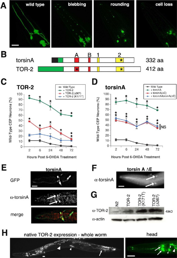
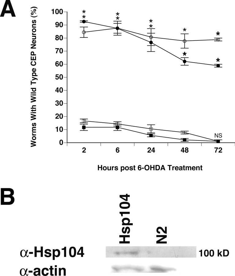
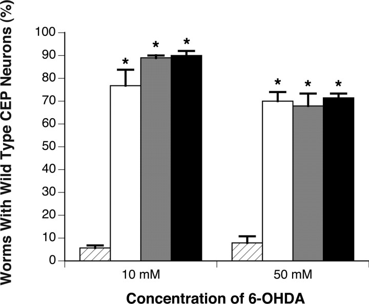
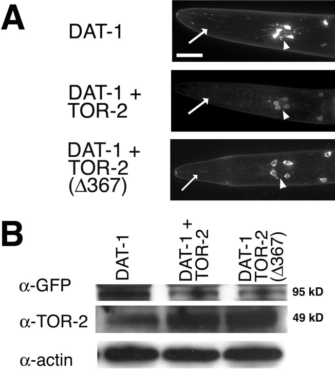
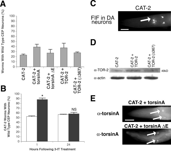
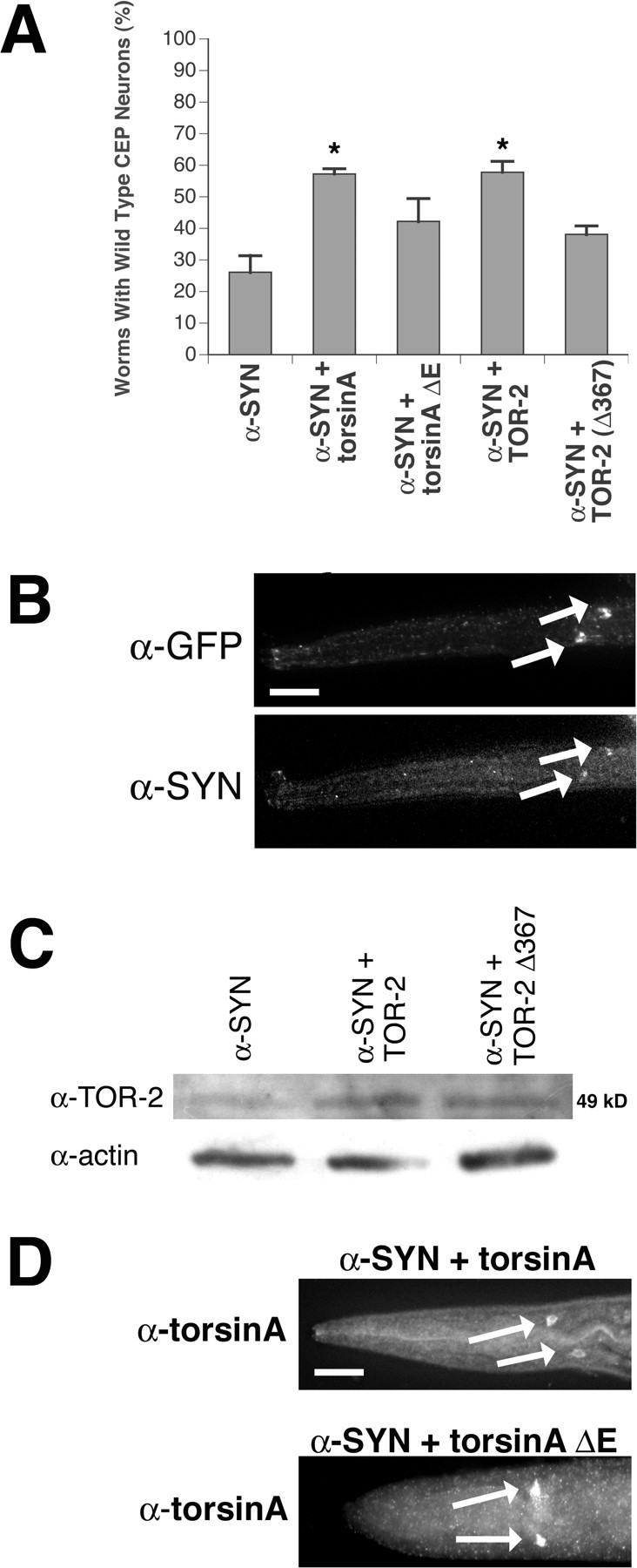
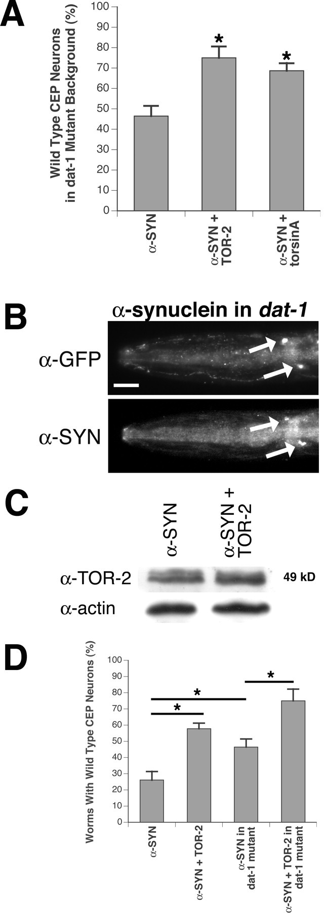
Parkinson's disease (PD) is linked genetically to proteins that function in the management of cellular stress resulting from protein misfolding and oxidative damage. Overexpression or mutation of alpha-synuclein results in the formation of Lewy bodies and neurodegeneration of dopaminergic (DA) neurons. Human torsinA, mutations in which cause another movement disorder termed early-onset torsion dystonia, is highly expressed in DA neurons and is also a component of Lewy bodies. Previous work has established torsins as having molecular chaperone activity. Thus, we examined the ability of torsinA to manage cellular stress within DA neurons of the nematode Caenorhabditis elegans. Worm DA neurons undergo a reproducible pattern of neurodegeneration after treatment with 6-hydroxydopamine (6-OHDA), a neurotoxin commonly used to model PD. Overexpression of torsins in C. elegans DA neurons results in dramatic suppression of neurodegeneration after 6-OHDA treatment. In contrast, expression of either dystonia-associated mutant torsinA or combined overexpression of wild-type and mutant torsinA yielded greatly diminished neuroprotection against 6-OHDA. We further demonstrated that torsins seem to protect DA neurons from 6-OHDA through downregulating protein levels of the dopamine transporter (DAT-1) in vivo. Additionally, we determined that torsins protect robustly against DA neurodegeneration caused by overexpression of alpha-synuclein. Using mutant nematodes lacking DAT-1 function, we also showed that torsin neuroprotection from alpha-synuclein-induced degeneration occurs in a manner independent of this transporter. Together, these data have mechanistic implications for movement disorders, because our results demonstrate that torsin proteins have the capacity to manage sources of cellular stress within DA neurons.


Figures

Figure 1.
Figure 1.
Torsin-mediated protection of C. elegans DA neurons against 6-OHDA. A, Different stages of DA neurodegeneration. From left to right, representative temporally staged images depicting CEP DA neuron morphology of wild type, neuronal process blebbing, process loss and cell body rounding, and cell body loss are shown. Scale bar, 20 μm. B, Schematic representation of torsin domains and mutations. Top, Human torsinA; bottom, C. elegans TOR-2; black region, signal peptide; green region, putative hydrophobic transmembrane domain; A, Walker A domain; B, Walker B domain; 1, sensor 1 domain; 2, sensor 2 domain. The asterisks mark the approximate positions of mutations that were analyzed. Mutations within sensor 2 correspond to the EOTD-associated mutation [torsinA(ΔE302/303)] and the structurally analogous change in worm TOR-2 [TOR-2(Δ367)]. The mutation within the Walker A domain corresponds to an alteration within a conserved ATP-binding site [TOR-2(K171T)]. C, TOR-2 protection against 6-OHDA. After 6-OHDA treatment, worms were examined for the presence of all four wild-type CEP neurons at specific time points: green, TOR-2; blue, TOR-2(K171T); red, TOR-2(Δ367); black, Pdat-1::GFP without TOR-2 overexpression. D, TorsinA protection against 6-OHDA. Green, TorsinA; blue, torsinA/torsinA (ΔE302/303) combination; red, torsinA(ΔE302/303); black, Pdat-1::GFP without torsinA overexpression. Statistical significance between Pdat-1::GFP and each of the six torsin-overexpressing lines was assessed at each time point between all transgenic lines by the ANOVA Bonferroni's test (*p < 0.05; NS, not significant). Error bars in C and D indicate SEM between independent experiments. E, Immunolocalization of torsinA in DA neurons in human torsin-overexpressing worms. Top, Two CEP neurons marked by GFP fluorescence; middle, torsinA antibody immunostaining in CEP cell bodies (arrows); bottom, merge of top and middle images. Scale bar, 20 μm. F, Immunolocalization of torsinA in torsinA(ΔE302/303)-overexpressing worms in CEP cell bodies (arrows). Scale bar, 20 μm. The GFP and merge images are not shown because they were reminiscent of those in Figure 1 E. G, Western analysis to verify TOR-2 overexpression in TOR-2-overexpressing worms. Actin was probed for each sample as a loading control. H, Native TOR-2 expression pattern in transgenic worms in which GFP is driven by a genomic region upstream and inclusive of the C. elegans tor-2 gene. Left, Entire worm depicting expression in the AVE neurons (large arrowhead), vulva muscle cells (small arrowhead), and PVW neurons (arrow); gut autofluorescence is also observed throughout the intestinal tract. Right, Close-up of the anterior head region, showing expression in the M1 pharyngeal neuron (large arrowhead), the AW class of neurons (2 small arrowheads), and the AVE interneurons (2 arrows). Expression in DA neurons is not observed. Scale bar, 20 μm.
Figure 7.
Figure 7.
Hsp104 does not protect against 6-OHDA-induced neurodegeneration. A, Comparison of the protective effects observed between torsins and yeast Hsp104 overexpressed in DA neurons. After 6-OHDA treatment, worms were examined for the presence of all four wild-type CEP neurons at specific time points. Filled circle, TOR-2; open circle, torsinA; open square, Hsp104; filled square, N2 background with only Pdat-1::GFP. Statistical significance between Pdat-1::GFP and each of the other transgenic lines was assessed by the ANOVA Bonferroni's test (*p < 0.05; NS, not significant). Error bars indicate SEM between independent experiments. B, Western analysis of Hsp104 expression in which Hsp104 was identified as a ∼100 kDa band present in extracts from Pdat-1::Hsp104 animals but not from N2 controls. Actin was probed as a loading control.
Figure 2.
Figure 2.
Transgenic animals overexpressing TOR-2 exhibit neuroprotection from 6-OHDA at levels comparable with dat-1 mutant worms. Twenty-four hours after exposure to either 10 or 50 mm 6-OHDA, Pdat-1::GFP control worms (hatched bars), TOR-2-overexpressing worms (white bars), Pdat-1::GFP in dat-1 background worms (gray bars), and TOR-2-overexpressing worms in the dat-1 mutant background (black bars) were scored for evidence of DA neuron survival (n = 30-40 worms per line per independent experiment). Statistical significance was determined between control and other transgenic lines by the ANOVA Bonferroni's test (*p < 0.05). Error bars indicate SEM between independent experiments.
Figure 3.
Figure 3.
TOR-2 downregulates GFP::DAT-1. A, GFP fluorescence is greatly diminished within the axons (arrowheads) and dendrites (arrows) of DA neurons when TOR-2 is co-overexpressed and moderately diminished when TOR-2(Δ367) is co-overexpressed with GFP::DAT-1. Scale bar, 20 μm. B, Western analysis of GFP::DAT-1 demonstrating that there is a higher level of DAT-1 in worms that do not co-overexpress TOR-2. GFP antibody was used to detect the GFP::DAT-1 fusion protein band; TOR-2 antibody was used to detect the level of TOR-2 expression. Actin was probed for each line as a loading control.
Figure 4.
Figure 4.
Torsin effect on TH/CAT-2 overexpression in DA neurons. A, A slight trend toward neuronal survival when torsinA or TOR-2 is coexpressed with CAT-2 is shown. The percentage of worms preserving all four wild-type CEPs at the 7-d-old stage was calculated for each of five CAT-2-overexpressing lines (n = 30-40 per stable line). Statistical significance between Pdat-1::CAT-2 and each of the torsin/CAT-2-overexpressing lines was assessed by the ANOVA Bonferroni's test; no significance was observed. B, 3-IT inhibition of CAT-2-induced neurodegeneration. Pdat-1::CAT-2 worms were incubated with or without 3-IT (black and white bars, respectively) in M9 buffer for 24 h, and neurodegeneration was analyzed 1 and 24 h after treatment. Statistical significance was assessed by t test (*p < 0.05; NS, not significant). Error bars in A and B indicate SEM between independent experiments. C, Strong FIF in CEP neuronal cell bodies (arrows) in Pdat-1::CAT-2 worms is indicative of high levels of dopamine. Scale bar, 20 μm. D, Western analysis of TOR-2 expression levels in TOR-2-overexpressing worms. Actin was probed as a loading control for each line. E, Immunolocalization of torsinA and torsinA(ΔE302/303) using anti-torsinA antibody in CEP cell bodies (arrows) of torsinA-overexpressing worms. Only torsinA immunostaining images are shown because GFP images were similar to those depicted in Figure 1 E. Scale bar, 20 μm.
Figure 5.
Figure 5.
Torsin-mediated protection of DA neurons against α-synuclein overexpression. A, Overexpression of torsinA or TOR-2 protects against α-synuclein overexpression. The percentage of worms preserving all four wild-type CEP neurons at the 7-d-old stage was calculated for each of five α-synuclein-overexpressing lines (n = 30-40 per stable line). Statistical significance between Pdat-1::α-synuclein and each of four torsin/α-synuclein-overexpressing lines was assessed by the ANOVA Bonferroni's test (*p < 0.05). Error bars indicate SEM between independent experiments. B, Immunolocalization of α-synuclein within CEP cell bodies (arrows) of α-synuclein-overexpressing worms by α-synuclein antibody. The position of CEP cell bodies is depicted with α-GFP antibody. This worm is representative of all α-synuclein-overexpressing lines. Merged images were similar to those in Figure 1E. Scale bar, 20 μm. C, Western analysis of TOR-2 expression levels in TOR-2-overexpressing worms. Actin was probed for each line as a loading control. D, Immunolocalization of torsinA and torsinA(ΔE302/303) by torsinA antibody in CEP cell bodies (arrows) of torsinA-overexpressing worms. Only torsinA immunostaining images are shown because GFP images were similar to those depicted in Figure 1E. Scale bar, 20 μm.
Figure 6.
Figure 6.
Torsin-mediated protection of DA neurons against α-synuclein overexpression in dat-1 knock-out animals. A, Overexpression of torsinA or TOR-2 protects against α-synuclein overexpression in dat-1 mutants. Percentage of 7-day-old worms preserving all four wild-type CEP neurons at each stage was calculated for each of three α-synuclein-overexpressing lines (n = 30-40 per stable line). Statistical significance between Pdat-1::α-synuclein and each of two α-synuclein/torsin-overexpressing lines was assessed by the ANOVA Bonferroni's test and is indicated with *p < 0.05. Error bars indicate SEM between independent experiments. B, Immunolocalization of α-synuclein within CEP cell bodies (arrows) of α-synuclein-overexpressing worms by α-synuclein antibody. The position of CEP cell bodies is depicted with GFP antibody. The worm depicted is representative of all α-synuclein-overexpressing lines. The merged images were similar to those in Figure 1 E. Scale bar, 20 μm. C, Western analysis of TOR-2 expression level in TOR-2-overexpressing worms. Actin was probed as a loading control. D, A comparison of DAT-1 depletion and torsin overexpression on α-synuclein-induced degeneration. Statistical significance was assessed by the ANOVA Bonferroni's test (*p < 0.05). Bars signify the worm strains compared in these analyses. Both overexpression of TOR-2 and loss of DAT-1 can contribute to neuroprotection against α-synuclein overexpression. Additionally, TOR-2 protection in DA neurons is independent of DAT-1. Error bars indicate SEM between independent experiments.

Similar articles

Cited by

References

    1. Augood SJ, Hollingworth Z, Albers DS, Yang L, Leung JC, Muller B, Klein C, Breakefield XO, Standaert DG (2002) Dopamine transmission in DYT1 dystonia: a biochemical and autoradiographical study. Neurology 59: 445-448. - PubMed
    1. Augood SJ, Hollingworth Z, Albers DS, Yang L, Leung J, Breakefield XO, Standaert DG (2004) Dopamine transmission in DYT1 dystonia. Adv Neurol 94: 53-60. - PubMed
    1. Auluck PK, Chan HY, Trojanowski JQ, Lee VM, Bonini NM (2002) Chaperone suppression of alpha-synuclein toxicity in a Drosophila model for Parkinson's disease. Science 295: 809-810. - PubMed
    1. Berger K, Przedborski S, Cadet JL (1991) Retrograde degeneration of nigrostriatal neurons induced by intrastriatal 6-hydroxydopamine injection in rats. Brain Res Bull 26: 301-307. - PubMed
    1. Blum D, Torch S, Lambeng N, Nissou M, Benabid AL, Sadoul R, Verna JM (2001) Molecular pathways involved in the neurotoxicity of 6-OHDA, dopamine and MPTP: contribution to the apoptotic theory in Parkinson's disease. Prog Neurobiol 65: 135-172. - PubMed

Publication types

MeSH terms

LinkOut - more resources