Comparative Study
. 2005 Apr 13;25(15):3900-7.
doi: 10.1523/JNEUROSCI.0102-05.2005.

Modular organization of excitatory circuits between neurons of the spinal superficial dorsal horn (laminae I and II)

Affiliations
Comparative Study

Modular organization of excitatory circuits between neurons of the spinal superficial dorsal horn (laminae I and II)

Yan Lu et al. J Neurosci. .

Abstract

Neural circuitry of the spinal superficial dorsal horn (SDH) (laminae I and II) and its relationship to pain and other somatosensory phenomena remain poorly understood. To gain information on this issue, synaptic connections between identified SDH neurons were studied in rat spinal cord slices by simultaneous whole-cell recordings from pairs of cells. Both excitatory and inhibitory connections were noted. This report focuses on the observed excitatory linkages. Synaptic excitatory connections between SDH neurons proved highly selective and consistently were unidirectional. Two patterns repeatedly appeared (for neuron classification, see Materials and Methods) (Grudt and Perl, 2002). Lamina II central neurons, with dorsal root (DR) C-fiber input, monosynaptically excited lamina II vertical neurons with DR Adelta input. Lamina II outer vertical neurons with DR Adelta input monosynaptically excited lamina I neurons. Some of the postsynaptic lamina I cells were shown to project rostrally. In contrast to the usual case for connected neurons, in unconnected pairs, primary afferent input to the same type of neuron proved closely similar. Together, these observations indicate that the neural circuitry in the SDH, including its substantia gelatinosa (lamina II), has an explicit organization in which particular combinations of neurons comprise modules arranged to modify and transmit sensory information arriving from Adelta and C primary afferent fibers.


Figures

Figure 1.
Figure 1.
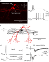
Examples of monosynaptic excitatory connections between SDH neurons. a, Lamina II transient central cell projection to lamina II vertical neuron. b, Lamina II vertical cell projecting to a lamina I neuron. Top traces, Five superimposed traces from the presynaptic neuron showing action potentials (Pre AP) initiated by depolarization pulses repeating at 0.2 Hz. Middle traces, Five traces of simultaneous recordings of EPSPs from the postsynaptic neuron (Post EPSPs). Bottom traces, Average of 50 successive EPSPs. A block of EPSPs by bath-applied CNQX (20 μm) is shown. The latencies from the peak of the presynaptic APs to onset of the EPSPs remain constant at 0.2 Hz (a) and in four repetitions at 70 ms intervals (b). The holding potential was -60 mV for both presynaptic and postsynaptic neurons.
Figure 2.
Figure 2.
Monosynaptic excitatory connection between a lamina II transient central neuron and a lamina II vertical neuron. a, Maximum projection confocal images of the biocytin-labeled connected neurons. Presynaptic, Lamina II transient central cell; postsynaptic, lamina II vertical cell. b, Action potential firing patterns of the connected neurons. c, Reconstructions of labeled neurons showing the presynaptic central cell in black and the postsynaptic vertical cell in red. Arrows indicate putative axons. Dotted lines mark approximate borders between dorsal horn laminae. d, Simultaneous whole-cell recordings illustrating the synaptic connection between the two neurons (average of 50 successive traces; membrane potential held at -60 mV for both cells). e, Simultaneous whole-cell recordings from the two neurons showing monosynaptic EPSCs evoked by dorsal root stimulation (holding potential, -60 mV for both cells). Five trials are superimposed. The lamina II transient central cell exhibits DR C-fiber input, whereas the lamina IIo vertical cell shows responses evoked by DR Aδ fibers. C, Caudal; D, dorsal; R, rostral; V, ventral; Pre, presynaptic; Post, postsynaptic; AP, action potential.
Figure 3.
Figure 3.
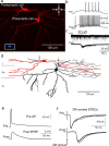
Monosynaptic excitatory connection from a lamina IIo vertical cell to a lamina I projection neuron. a, Maximum projection confocal images of the connected cells. Presynaptic, Lamina IIo vertical neuron; postsynaptic, lamina I neuron. The bottom left insert shows retrogradely transported FG from the thoracic spinal cord labeling the lamina I cell. b, Action potential firing patterns of the connected neurons. c, Simultaneous voltage-clamp recordings showing inward current in the lamina I cell evoked by substance P (2 μm) in the presence of 0.5 μm TTX but not in the lamina II vertical cell. d, Reconstructions of the labeled neurons depicting the presynaptic lamina IIo vertical cell in black and the postsynaptic lamina I cell in red. Arrows indicate putative axons. e, Simultaneous current-clamp recordings showing the synaptic connections between two neurons (average of 50 successive traces; membrane potential, -60 mV for both cells). f, Simultaneous voltage-clamp recordings from the pair showing monosynaptic EPSCs evoked by dorsal root stimulation (holding potential, -60 mV for both cells). Five trials are superimposed. The vertical cell has a monosynaptic dorsal root Aδ-fiber input; the lamina I projection cell receives monosynaptic DR C-fiber connections. C, Caudal; D, dorsal; R, rostral; V, ventral; Pre, presynaptic; Post, postsynaptic; AP, action potential.
Figure 4.
Figure 4.
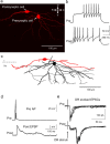
Monosynaptic excitatory linkage from a lamina IIo vertical cell to a lamina I, substance P-insensitive neuron. a, Maximum projection confocal images of the connected cells. Presynaptic, Lamina IIo vertical cell. Postsynaptic, Lamina I neuron. b, Action potential firing patterns of the connected neurons. c, Reconstructions of the labeled neurons showing the presynaptic lamina IIo neuron in black and the postsynaptic lamina I cell in red. Arrows indicate putative axons.d, Simultaneous current-clamp recording showing the synaptic connection (average of 50 successive traces; membrane potential, -60 mV for both cells). e, Simultaneous voltage-clamp recordings depicting the monosynaptic EPSCs evoked by dorsal root stimulation in the pair (holding potential, -60 mV for both cells). Five trials are superimposed. The vertical neuron receives monosynaptic DR Aδ input, whereas the lamina I, substance P-insensitive neuron shows both monosynaptic Aδ- and C-fiber DR input. C, Caudal; D, dorsal; R, rostral; V, ventral; Pre, presynaptic; Post, postsynaptic; AP, action potential.
Figure 5.
Figure 5.
Simultaneously recorded, unconnected lamina II vertical neurons receiving closely similar DR Aδ-fiber input. a, Maximum projection confocal image of the two biocytin-labeled neurons. b, Action potential firing patterns of the two vertical neurons. c, Reconstructions of labeled neurons: one vertical cell is black, and the other is red. Arrows indicate putative axons. d, Simultaneous voltage-clamp recordings showing monosynaptic EPSCs evoked by dorsal root stimulation (holding potential, -60 mV for each cell). Five trials are superimposed. Both cells receive monosynaptic excitatory input from dorsal root Aδ fibers with the same or similar thresholds. Note the identical latencies and threshold stimulus intensities for the two cells. C, Caudal; D, dorsal; R, rostral; V, ventral.
Figure 6.
Figure 6.
Simultaneously recorded, unconnected lamina II transient central cells receiving closely similar DR C-fiber input. a, Photomicrograph of the two biocytin-labeled transient central cells. b, Action potential firing pattern of the two transient central neurons.c, Reconstructions of labeled neurons; one central cell is shown in black, the other in red. The arrow indicates the putative axon of the black cell. d, Simultaneous voltage-clamp recordings showing the monosynaptic EPSCs evoked by dorsal root stimulation in the two cells (holding potential, -60 mV for each cell). Five trials are superimposed. Both cells receive monosynaptic excitatory input from same or similar threshold dorsal root C fibers. Note the identical latencies and stimulus intensities generating the EPSCs in the two neurons. C, Caudal; D, dorsal; R, rostral; V, ventral.
Figure 7.
Figure 7.
Schematic summarizing excitatory circuitry in the SDH established by the present study. Certain lamina II transient central neurons have a monosynaptic glutamatergic (Glu) connection to lamina IIo vertical neurons. Certain lamina IIo vertical neurons make monosynaptic glutamatergic projections to lamina I neurons, including some that possess substance P receptors and contribute to rostrally directed pathways. All of these cells have monosynaptic, AMPA receptor-mediated input from primary afferents. Vertical cells receive DR Aδ-fiber input, whereas most lamina I and lamina II central neurons have direct connections from DR C fibers. The schematic does not show an Aδ primary afferent projection to lamina I neurons because this was an inconsistent feature. This circuitry is interpreted as representing modules capable of amplifying and manipulating the primary afferent input from DR Aδ and C fibers in the process of forwarding signals to other CNS centers. DRG, Dorsal root ganglion; C, caudal; D, dorsal; R, rostral; V, ventral.

References

    1. Bennett GJ, Abdelmoumene M, Hayashi H, Dubner R (1980) Physiology and morphology of substantia gelatinosa neurons intracellularly stained with horseradish peroxidase. J Comp Neurol 194: 809-827. - PubMed
    1. Craig AD (1994) Spinal and supraspinal processing of specific pain and temperature. In: Touch, temperature, and pain in health and disease: mechanisms and assessments (Boivie J, Hansson P, Lindblom U, eds), pp 421-437. Seattle: International Association for the Study of Pain.
    1. Craig AD, Kniffki KD (1985) The multiple representation of nociception in the spinothalamic projection of lamina I cells in the cat. In: Development, organization, and processing in somatosensory pathways (Rowe M, Willis W, eds), pp 347-353. New York: Alan R. Liss.
    1. Earle KM (1952) The tract of Lissauer and its possible relation to the pain pathway. J Comp Neurol 96: 93-109. - PubMed
    1. Gobel S (1978) Golgi studies of the neurons in layer II of the dorsal horn of the medulla (trigeminal nucleus caudalis). J Comp Neurol 180: 395-414. - PubMed

Publication types

MeSH terms

LinkOut - more resources