

The suprapedicle claw construct in anterior scoliosis surgery
- PMID: 15830213
- PMCID: PMC3489252
- DOI: 10.1007/s00586-004-0804-3
The suprapedicle claw construct in anterior scoliosis surgery
Abstract
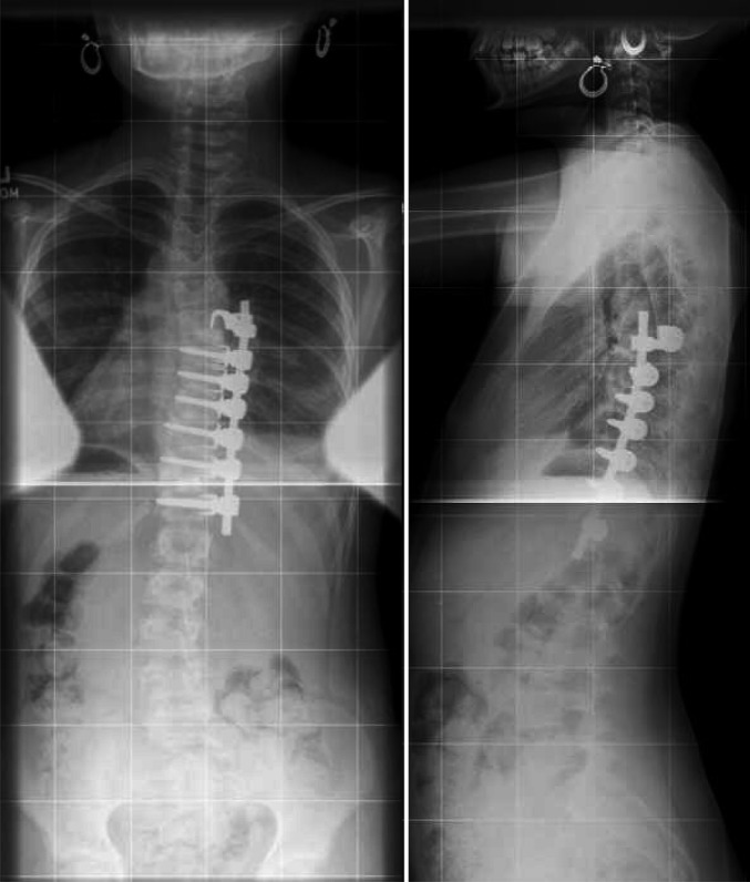
Proximal screw pullout is a well-recognized problem in anterior scoliosis surgery, with a rate of pseudarthrosis or screw pullout ranging from 15 to 30%. To prevent screw pullout at the top of the construct, the authors have devised the concept of a claw for the top instrumented vertebra. The claw consists of a classic anterior vertebral body screw inserted parallel to the inferior end-plate and in the posterior portion of the vertebral body 8 mm in front of the spine canal. After rib desarticulation, a laminar hook of a small size is inserted over the superior aspect of the pedicle of the same vertebra. The rod is then inserted into the two side openings of the screw and the hook. Compression across the hook and the screw is then performed, making a claw construct. This concept can also be extended in the case of early revision for a proximal screw pullout, where it is possible to revise the instrumentation with an offset connector linking the rod to the superior portion of the pedicle where the suprapedicule hook has been inserted. We report two cases where a suprapedicle claw was successfully used in anterior scoliosis correction of a right thoracic curve. Such a concept may represent the solution to proximal screw pullout in anterior scoliosis correction.
Figures


Similar articles
-
Pull-out strength of the suprapedicle claw construct: a biomechanical study.Eur Spine J. 2005 Oct;14(8):759-64. doi: 10.1007/s00586-004-0805-2. Epub 2005 Apr 14. Eur Spine J. 2005. PMID: 15830212 Free PMC article.
-
Comparative analysis of pedicle screw and hook instrumentation in posterior correction and fusion of idiopathic thoracic scoliosis.Eur Spine J. 2002 Aug;11(4):336-43. doi: 10.1007/s00586-002-0415-9. Epub 2002 May 29. Eur Spine J. 2002. PMID: 12193995 Free PMC article.
-
Analysis of screw placement relative to the aorta and spinal canal following anterior instrumentation for thoracic idiopathic scoliosis.Spine (Phila Pa 1976). 2004 Mar 1;29(5):554-9; discussion 559. doi: 10.1097/01.brs.0000106495.91477.92. Spine (Phila Pa 1976). 2004. PMID: 15129071
-
Comparative analysis of pedicle screw versus hook instrumentation in posterior spinal fusion of adolescent idiopathic scoliosis.Spine (Phila Pa 1976). 2004 Sep 15;29(18):2040-8. doi: 10.1097/01.brs.0000138268.12324.1a. Spine (Phila Pa 1976). 2004. PMID: 15371706 Review.
-
Pedicle screw instrumentation in adolescent idiopathic scoliosis (AIS).Eur Spine J. 2012 Jan;21(1):13-22. doi: 10.1007/s00586-011-1986-0. Epub 2011 Aug 30. Eur Spine J. 2012. PMID: 21874625 Free PMC article. Review.
Cited by
-
Pull-out strength of the suprapedicle claw construct: a biomechanical study.Eur Spine J. 2005 Oct;14(8):759-64. doi: 10.1007/s00586-004-0805-2. Epub 2005 Apr 14. Eur Spine J. 2005. PMID: 15830212 Free PMC article.
References
-
- Aebi M, Thalgott JS, Webb JK. AOASIF principles in spine surgery. Scoliosis anterior correction and stabilisation. Berlin Heidelberg New York: Springer; 1998.
-
- Betz RR, Shufflebarger H. Anterior versus posterior instrumentation for the correction of thoracic idiopathic scoliosis. Spine. 2001;26:1095–1100. - PubMed
-
- Gertzbein Spine. 1990;15:11. - PubMed
MeSH terms
LinkOut - more resources
Full Text Sources
Medical
