Review
. 2005 Apr;7(2):134-53.
doi: 10.1215/S1152851704001115.

Hypoxia and the hypoxia-inducible-factor pathway in glioma growth and angiogenesis

Affiliations
Review

Hypoxia and the hypoxia-inducible-factor pathway in glioma growth and angiogenesis

Balveen Kaur et al. Neuro Oncol. 2005 Apr.

Abstract

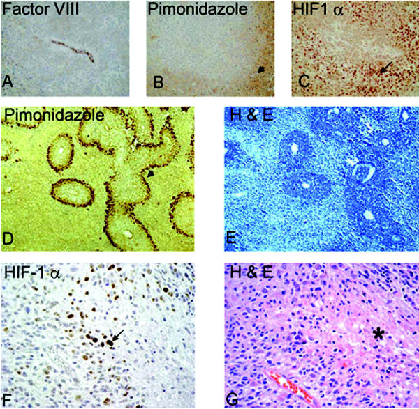
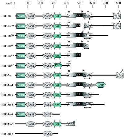
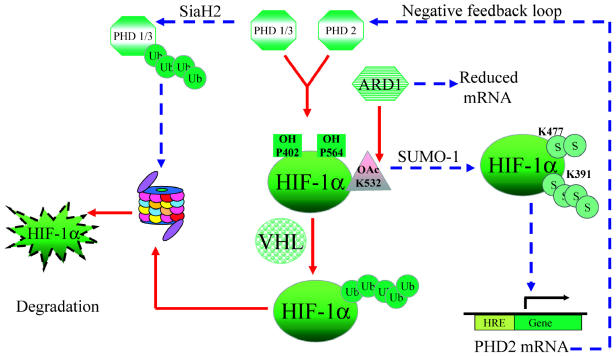
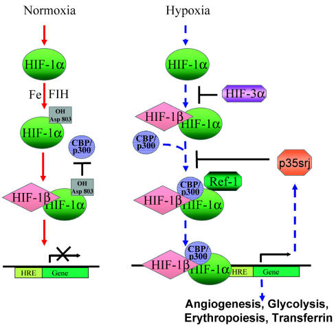
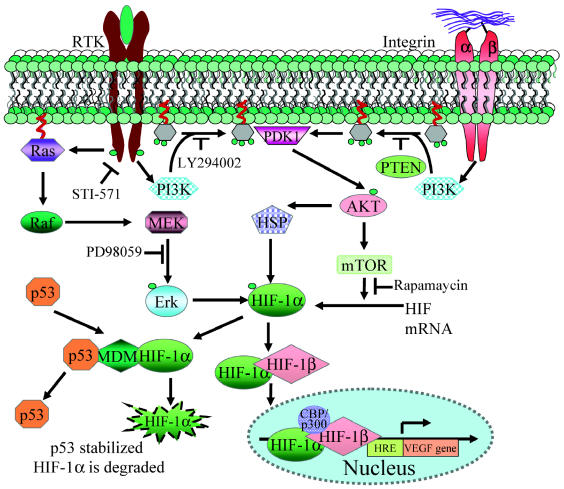
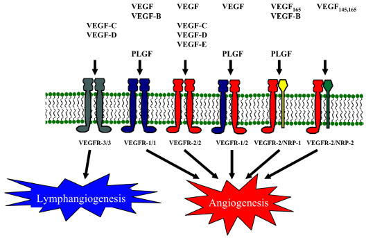
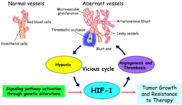
Glioblastomas, like other solid tumors, have extensive areas of hypoxia and necrosis. The importance of hypoxia in driving tumor growth is receiving increased attention. Hypoxia-inducible factor 1 (HIF-1) is one of the master regulators that orchestrate the cellular responses to hypoxia. It is a heterodimeric transcription factor composed of alpha and beta subunits. The alpha subunit is stable in hypoxic conditions but is rapidly degraded in normoxia. The function of HIF-1 is also modulated by several molecular mechanisms that regulate its synthesis, degradation, and transcriptional activity. Upon stabilization or activation, HIF-1 translocates to the nucleus and induces transcription of its downstream target genes. Most important to gliomagenesis, HIF-1 is a potent activator of angiogenesis and invasion through its upregulation of target genes critical for these functions. Activation of the HIF-1 pathway is a common feature of gliomas and may explain the intense vascular hyperplasia often seen in glioblastoma multiforme. Activation of HIF results in the activation of vascular endothelial growth factors, vascular endothelial growth factor receptors, matrix metalloproteinases, plasminogen activator inhibitor, transforming growth factors alpha and beta, angiopoietin and Tie receptors, endothelin-1, inducible nitric oxide synthase, adrenomedullin, and erythropoietin, which all affect glioma angiogenesis. In conclusion, HIF is a critical regulatory factor in the tumor microenvironment because of its central role in promoting proangiogenic and invasive properties. While HIF activation strongly promotes angiogenesis, the emerging vasculature is often abnormal, leading to a vicious cycle that causes further hypoxia and HIF upregulation.


Figures

Fig. 1
Fig. 1
HIF stabilization in hypoxic areas. HIF-1α is stabilized in cells distant from a blood vessel. A–C: Adjacent sections of subcutaneously grown tumor xenografts of human LN229 glioma cells. Panel A shows stain Factor VIII, highlighting a vessel. Panel B is pimonidazole staining showing hypoxic areas in brown (arrowhead). Panel C is HIF-1α immunostain (arrow), showing nuclear staining of HIF. D–E: U87 glioma xenografts from a rat orthotopic brain tumor model. Panel D is pimonidazole staining that shows a rim of viable hypoxic cells at the periphery of vascularized regions (arrowhead). Panel E shows the corresponding H & E staining. Note large areas of necrosis (light blue). F–G: Human GBM specimen. Panel F highlights the HIF-1α-positive staining cells localized in the pseudopalisading cells (arrow), and Panel G is the corresponding H & E of the adjacent section showing the necrotic area (asterisk).
Fig. 2
Fig. 2
Domain structure of HIF-1α1–6 (shown as HIF-1α–HIF-1α417), HIF-2α, and HIF-3α1–6. Functional domains are abbreviated as follows: bHLH, basic helix-loop-helix; PAS, Per/Arnt/Sim; PAC, PAS-associated motifs; NAD, N-terminal transactivation domain; ODD, oxygen-dependent domain; CAD, C-terminal activation domain; and LZIP, leucine zipper domain. Position of modified amino acids are indicated as follows: *, hydroxylated proline; @, acetylated lysine;Δ, hydroxylated asparagines. (Adapted from Maynard et al. [2003] and Lee et al. [2004].)
Fig. 3
Fig. 3
Factors affecting HIF-1α protein stability. PHD-mediated hydroxylations and ARD-mediated acetylation of specific residues within HIF-1α increase its affinity for pVHL, which leads to its ubiquitination (Ub) and degradation by the proteasomal pathway under normoxia (solid arrows). PHD1, 2, and 3 have a reduced catalytic activity in the absence of oxygen. Further, PHD1 and 3 and ARD have reduced levels in hypoxia (dashed arrows), adding another level of control. SUMO-1-mediated sumoylation in hypoxia leads to HIF-1α stabilization (S) and activation, causing transactivation of specific downstream target genes. PHD2 is induced by HIF, which indicates a negative feedback loop.
Fig. 4
Fig. 4
HIF-1α is differentially regulated under normoxia versus hypoxia. In normoxia (solid arrows), hydroxylation of HIF-1α mediated by asparaginyl hydroxylase FIH-1 interferes with its ability to bind co-activator CBP/p300, which is necessary to form an active HIF complex. Under hypoxia (dashed arrows), HIF-1α is stabilized and translocates to the nucleus after binding to HIF-1β, where Ref-1 aids in the recruitment of CBP/p300 to the HIF-1α complex, leading to transcriptional activation of genes containing the HREs. CITED2/p35srj negatively regulates HIF activity by competing with HIF-1α for binding to the CH1 region of CBP/p300. It is also upregulated by HIF, which indicates a possible negative feedback regulation.
Fig. 5
Fig. 5
Molecular signals affecting HIF-1α regulation. Induction of Ras, PI3K, and AKT phosphorylation mediated by RTK activation or integrin ligation leads to increased HIF-1α by modulating its stability and increased translation by the PI3K/AKT/mTOR pathway. TP53 negatively modulates this process by inducing MDM2, which can ubiquitinate and lead to HIF-1α degradation by the proteasome pathway.
Fig. 6
Fig. 6
VEGF and VEGF-receptor interactions. The various family members of VEGF bind differentially to a variety of receptors. The VEGFR-1 (Flt-1) and VEGFR-2 (Flk-1/KDR) have been predominately found in endothelial cells, but expression has also been reported in glioma tissues and glioma cell lines. VEGFR-3 is mainly expressed on the endothelial cells in the lymphatics and is thought to be involved in lymphangiogenesis. Both NRP-1 and NRP-2 are neuronal and endothelial cell surface glycoproteins that dimerize with VEGFR-2 and are expressed in some GBMs and to a lesser extent in low-grade astrocytomas.
Fig. 7
Fig. 7
Vicious HIF activation cycle in glioma. Genetic alterations (such as loss of PTEN or EGFR mutation) affect critical signaling pathways that lead to HIF-1 stabilization and activation. Transcriptional activity by HIF-1 directly upregulates genes that promote angiogenesis, such as VEGF. Although the upregulation and secretion of proangiogenic factors by tumor cells leads to a strong angiogenic response, the resulting tumor vasculature is abnormal, displaying leakiness due to excessive vascular permeability, focal thickening of vascular walls, and the formation of glomeruloids arising from excessive microvascular proliferation. The tumor vasculature also exhibits abnormal vessel branching, arteriovenous shunts, poor blood flow, and low structural integrity due to inappropriate strengthening of nascent vessels by smooth muscle/pericyte lining. Thrombotic events that arise from poor blood flow and increased tissue factor expression by the neoplasm lead to vascular occlusion. Combined, the vascular phenomena lead to further escalating levels of hypoxia, which in turn stimulates more HIF-1 expression and activation. These events take place at the growing edge of the tumor and lead to a constellation of microregions of hypoxia that develop into pseudopalisading necrosis fueling further HIF expression, angiogenesis, and peripheral tumor expansion. All of these factors lead to tumor growth and resistance to therapy, with HIF-1 as a main component of this vicious cycle.

References

    1. Appelhoff RJ, Tian YM, Raval RR, Turley H, Harris AL, Pugh CW, Ratcliffe PJ, Gleadle JM. Differential function of the prolyl hydroxylases PHD1, PHD2, and PHD3 in the regulation of hypoxia-inducible factor. J Biol Chem. 2004;279:38458–38465. - PubMed
    1. Asano T, Aoyagi M, Hirakawa K, Ikawa Y. Effect of endothelin-1 as growth factor on a human glioma cell line; its characteristic promotion of DNA synthesis. J Neurooncol. 1994;18:1–7. - PubMed
    1. Bagnato A, Spinella F. Emerging role of endothelin-1 in tumor angiogenesis. Trends Endocrinol Metab. 2003;14:44–50. - PubMed
    1. Bajou K, Maillard C, Jost M, Lijnen RH, Gils A, Declerck P, Carmeliet P, Foidart JM, Noel A. Host-derived plasminogen activator inhibitor-1 (PAI-1) concentration is critical for in vivo tumoral angiogenesis and growth. Oncogene. 2004;23:6986–6990. - PubMed
    1. Barker FG, 2nd, Davis RL, Chang SM, Prados MD. Necrosis as a prognostic factor in glioblastoma multiforme. Cancer. 1996;77:1161–1166. - PubMed

Publication types