. 2005 May;25(9):3443-51.
doi: 10.1128/MCB.25.9.3443-3451.2005.

Adult stage gamma-globin silencing is mediated by a promoter direct repeat element

Affiliations

Adult stage gamma-globin silencing is mediated by a promoter direct repeat element

Akane Omori et al. Mol Cell Biol. 2005 May.

Abstract

The human beta-like globin genes (5'-epsilon-Ggamma-Agamma-delta-beta-3') are temporally expressed in sequential order from the 5' to 3' end of the locus, but the nonadult epsilon- and gamma-globin genes are autonomously silenced in adult erythroid cells. Two cis elements have been proposed to regulate definitive erythroid gamma-globin repression: the DR (direct repeat) and CCTTG elements. Since these two elements partially overlap, and since a well-characterized HPFH point mutation maps to an overlapping nucleotide, it is not clear if both or only one of the two participate in gamma-globin silencing. To evaluate the contribution of these hypothetical silencers to gamma-globin regulation, we generated point mutations that individually disrupted either the single DR or all four CCTTG elements. These two were separately incorporated into human beta-globin yeast artificial chromosomes, which were then used to generate gamma-globin mutant transgenic mice. While DR element mutation led to a dramatic increase in Agamma-globin expression only during definitive erythropoiesis, the CCTTG mutation did not affect adult stage transcription. These results demonstrate that the DR sequence element autonomously mediates definitive stage-specific gamma-globin gene silencing.


Figures
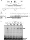
FIG. 1.
FIG. 1.
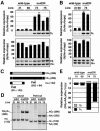
Human Aγ-globin gene promoter mutagenesis. (A) Schematic structure of the human β-globin locus used for creating transgenic mice. The LCR and β-like globin genes are shown by the open and solid boxes, respectively (top). An enlarged map of Aγ-globin gene promoter is shown (middle) with two putative repressor sequences, a DR (shaded arrow) motif and four CCTTG motifs (open rectangles). The two motifs partially overlap, and the Greek-type HPFH mutation (Aγ-117, indicated as a vertical line) maps in this overlapping segment. Detailed sequences of the Aγ promoter are shown (bottom) with a CAC and distal CAAT boxes (bracketed). The DR motif is underlined with CCTTG motifs italicized. (B) Sequence alignment of wild-type (WT) and mutant Aγ-globin promoters (mutDR and mutCCTTG) used for YAC mutagenesis as well as the ɛ- and β-globin promoters (Epsi and Beta, respectively). These are aligned with DR motifs underlined, except for Beta, which has no DR motif (corresponding portions are still underlined). The CCTTG motifs are italicized and mutated nucleotides are indicated by periods. (C) Competitive EMSA analysis of DRED binding to the β-like globin gene promoters. Increasing amounts of MEL cell nuclear extract (NE) were incubated with [γ-32P]-labeled Epsi probe and analyzed by polyacrylamide gel electrophoresis. An unlabeled 50- or 200-fold molar excess of competitor oligonucleotides listed in panel B were included in the reaction. The relative abundance of DRED EMSA product is shown at the bottom of each lane (the signal intensity with no added competitor was set at 100%). −, no added MEL cell NE.
FIG. 2.
FIG. 2.
Structural analysis of human β-globin YAC TgM. (A) Schematic representation of the human β-globin YAC. The positions of the β-like globin genes are shown relative to the LCR. SfiI restriction enzyme sites are indicated as vertical lines. Probes (hatched boxes) used for long-range structural analysis and anticipated restriction enzyme fragments after SfiI digestion are shown with their sizes (solid thick lines). (B) Long-range transgene analysis of mutDR (left panel) and mutCCTTG (right panel) YAC transgenic mice. The whole β-globin locus is contained within two SfiI fragments (10 and 100 kb, as in panel A). DNA from thymus cells of transgenic mice was digested with SfiI in agarose plugs, separated by pulsed-field gel electrophoresis, and hybridized separately to probes (indicated on the top of each panel) from the β-globin locus or the right YAC vector arm. The sizes of the expected bands are shown on the left.
FIG. 3.
FIG. 3.
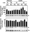
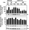
Expression of human β-like globin genes in the adult spleens of mutDR YAC TgM. (A) Semiquantitative RT-PCR analysis of β-like globin gene expression. Total RNA from two individuals from each line of TgM was prepared from the spleens of 1-month-old anemic mice. Expression of human γ (hγ)- and human β (hβ)-globin in comparison to the endogenous mouse α (mα)-globin genes was analyzed separately by semiquantitative RT-PCR. The signals for hγ-globin at 21 cycles and hβ/mα-globin at 12 cycles were quantified by PhosphorImager, and the ratios of hγ/mα (top) and hβ/mα (bottom) were calculated (the mouse α signal at 12 cycles was set at 100%, and the values are normalized by transgene copy numbers) and statistically analyzed (n = 3). The average and standard deviation are graphically depicted. Representative results are shown below each panel. (B) Total RNA from the peripheral blood of TgM was analyzed as in panel A. The PCR cycle numbers used were 20 and 11 for hγ-globin and hβ/mα-globin, respectively. (C to E) RT-PCR analysis of human Gγ/Aγ transcription ratios in the adult spleen. (C) Schematic representation of RT-PCR products amplified with primer sets specific for human γ- (common to both human Gγ- and Aγ-) and mouse α-globin genes. The positions of the primers (solid box), the total length of each RT-PCR product (hatched box for human Gγ, solid box for human Aγ, and open box for mouse α; sizes in base pairs are indicated in parentheses), and the positions of PstI restriction sites with the fragment sizes produced after enzyme digestion are shown in parentheses. (D) PstI digestion of RT-PCR products. The PCR products were digested with PstI and separated on an 8% polyacrylamide gel. A PstI site in the Aγ but not in the Gγ gene enabled separation and quantification of products derived from the two individual γ-globin genes. To internally control for complete PstI digestion, a PstI site was artificially introduced into the mouse α-globin gene PCR primer. The sizes of the expected bands are shown (on the left for undigested PCR products, and on the right for those digested with PstI; sizes in base pairs are indicated in parentheses). (E) The contribution of the Gγ and Aγ genes relative to total γ-globin synthesis are quantified from panel D and plotted as hatched and solid bars, respectively. Average values from two independent experiments are shown.
FIG. 4.
FIG. 4.
Expression of human β-like globin genes in the fetal liver of mutDR TgM. (A) Semiquantitative RT-PCR analysis of β-like globin gene expression. Total RNA was prepared from the livers of two fetuses (14.5 dpc) from two independent litters for each line, derived from the intercross of male transgenic and female wild-type animals. Expression of human γ (hγ)- and human β (hβ)-globin to endogenous mouse α (mα)-globin genes was analyzed separately by semiquantitative RT-PCR. The signals for hγ-globin at 18 cycles and hβ/mα-globin at 12 cycles were quantified by PhosphorImager, and the ratios of hγ/mα (top) and hβ/mα (bottom) were calculated (the mouse α signal at 12 cycles was set at 100% and the values are normalized by transgene copy numbers). The averages ± standard deviations from at least three independent experiments were calculated and are graphically depicted. Representative results are shown below each panel. (B and C) RT-PCR analysis of human Gγ/Aγ transcription ratios in the fetal liver. (B) PstI digestion of RT-PCR products. See the legend to Fig. 3 for details. Undigested PCR products are included (uncut) as a reference. (C) The contribution of the Gγ and Aγ genes relative to total γ-globin synthesis are quantified from panel B and plotted as hatched and solid bars, respectively. Average values obtained from two independent experiments are shown.
FIG. 5.
FIG. 5.
Expression of human β-like globin genes in the yolk sac of mutDR TgM. Semiquantitative RT-PCR analysis of β-like globin gene expression. Total RNA was prepared from the yolk sac of two embryos (9.5 dpc) derived from the intercross of male transgenic and female wild-type animals. Samples were collected from two independent litters from each mutant line. Expression of human ɛ (hɛ)- and human γ (hγ)-globin compared to endogenous mouse α (mα)-globin genes was analyzed separately by semiquantitative RT-PCR. The signals for hɛ-globin at 18 cycles and hγ/mα-globin at 12 cycles were quantified by PhosphorImager, and the ratios of hɛ/mα (top) and hγ/mα (bottom) were calculated (the mouse α signal at 12 cycles was set at 100% and the values are normalized by transgene copy numbers). The average ± standard deviation from at least three independent experiments was calculated and graphically depicted. Representative results are shown below each panel.
FIG. 6.
FIG. 6.
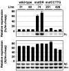
Expression of human β-like globin genes in the adult spleens of mutCCTTG TgM. Semiquantitative RT-PCR analysis of β-like globin gene expression. Total RNA from two individuals for each line was prepared from the spleens of anemic mice (1 month old). The average and standard deviation from three independent experiments are graphically depicted. Representative results are shown below each panel. See the legend to Fig. 3 for details.
FIG. 7.
FIG. 7.
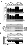
Expression of human β-like globin genes in the fetal liver of mutCCTTG TgM. (A) Semiquantitative RT-PCR analysis of β-like globin gene expression. Total RNA was prepared from the livers of two fetuses (14.5 dpc) in two independent litters (lit.1 and lit.2) for each line. The average and standard deviation from at least three independent experiments are graphically depicted. Representative results are shown below each panel. See the legend to Fig. 4 for details. (B and C) RT-PCR analysis of human Gγ/Aγ transcription ratios in the fetal liver. (B) PstI digestion of RT-PCR products. See the legend to Fig. 3 for details. (C) The contribution of the Gγ and Aγ genes relative to total γ-globin synthesis are quantified from panel B and plotted as hatched and solid bars, respectively. Average values obtained from two independent experiments are shown.
FIG. 8.
FIG. 8.
Expression of human β-like globin genes in the yolk sac of mutCCTTG TgM. Semiquantitative RT-PCR analysis of β-like globin gene expression. Total RNA was prepared from the yolk sac of two embryos (9.5 dpc) derived from the intercross of male transgenic and female wild-type animals. Samples were collected from two independent litters (lit.1 and lit.2) for each mutant line. The average ± standard deviation was calculated (n = 3) and is graphically depicted. Representative results are shown below each panel. See the legend to Fig. 5 for details.

Similar articles

Cited by

References

    1. Asano, H., and G. Stamatoyannopoulos. 1998. Activation of beta-globin promoter by erythroid Kruppel-like factor. Mol. Cell. Biol. 18:102-109. - PMC - PubMed
    1. Ashe, H. L., J. Monks, M. Wijgerde, P. Fraser, and N. J. Proudfoot. 1997. Intergenic transcription and transinduction of the human beta-globin locus. Genes Dev. 11:2494-2509. - PMC - PubMed
    1. Bungert, J., U. Dave, K. C. Lim, K. H. Lieuw, J. A. Shavit, Q. Liu, and J. D. Engel. 1995. Synergistic regulation of human beta-globin gene switching by locus control region elements HS3 and HS4. Genes Dev. 9:3083-3096. - PubMed
    1. Clouston, W. M., B. A. Evans, J. Haralambidis, and R. I. Richards. 1988. Molecular cloning of the mouse angiotensinogen gene. Genomics 2:240-248. - PubMed
    1. Collins, F. S., J. E. Metherall, M. Yamakawa, J. Pan, S. M. Weissman, and B. G. Forget. 1985. A point mutation in the A gamma-globin gene promoter in Greek hereditary persistence of fetal haemoglobin. Nature 313:325-326. - PubMed

Publication types

MeSH terms

LinkOut - more resources