. 2005 May;25(9):3492-505.
doi: 10.1128/MCB.25.9.3492-3505.2005.

Orphan nuclear receptor LRH-1 is required to maintain Oct4 expression at the epiblast stage of embryonic development

Affiliations

Orphan nuclear receptor LRH-1 is required to maintain Oct4 expression at the epiblast stage of embryonic development

Peili Gu et al. Mol Cell Biol. 2005 May.

Abstract

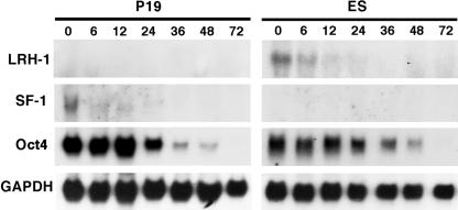
Oct4 plays an essential role in maintaining the inner cell mass and pluripotence of embryonic stem (ES) cells. The expression of Oct4 is regulated by the proximal enhancer and promoter in the epiblast and by the distal enhancer and promoter at all other stages in the pluripotent cell lineage. Here we report that the orphan nuclear receptor LRH-1, which is expressed in undifferentiated ES cells, can bind to SF-1 response elements in the proximal promoter and proximal enhancer of the Oct4 gene and activate Oct4 reporter gene expression. LRH-1 is colocalized with Oct4 in the inner cell mass and the epiblast of embryos at early developmental stages. Disruption of the LRH-1 gene results in loss of Oct4 expression at the epiblast stage and early embryonic death. Using LRH-1(-/-) ES cells, we also show that LRH-1 is required to maintain Oct4 expression at early differentiation time points. In vitro and in vivo results show that LRH-1 plays an essential role in the maintenance of Oct4 expression in ES cells at the epiblast stage of embryonic development, thereby maintaining pluripotence at this crucial developmental stage prior to segregation of the primordial germ cell lineage at gastrulation.


Figures

FIG. 1.
FIG. 1.
Northern blot analysis of SF1, LRH-1, and Oct4 expression in P19 and ES cells at different time points of RA-induced differentiation. The numbers above the figures indicate the RA (1 μM) treatment time (hours). Glyceraldehyde-3-phosphate dehydrogenase (GAPDH) is an internal control for loading and RNA integrity.
FIG. 2.
FIG. 2.
Direct binding of SF-1/LRH-1 to the Oct4 proximal enhancer and promoter. (A) Localization of the LRH-1- binding sites, PE1, PE2, and DR0 elements in the Oct4 proximal enhancer and promoter. The SF-1/LRH-1-binding sites were identified by HMMER and labeled as black bars in the mouse Oct4 gene. The relative conservation between the mouse and human Oct4 gene derived from the UCSC genome browser was plotted as the gray curve. Arrows indicate the three regulatory regions DE, PE, and PP. The highly conserved regions CR1, 2, 3 and 4 are depicted as open boxes. The LRH-1- binding sites PE1, PE2, and DR0 that were tested are shown as black bars in CR2 and CR1. (B) COS1-overexpressed SF-1 and LRH-1 binding to the PE1, PE2, and DR0 probes in the absence or presence of antibodies. (C) Total proteins extracted from undifferentiated or differentiated ES cells were incubated with DR0 probe in the absence or presence of LRH-1 or SF-1 antibodies. The arrowheads mark the SF-1- and LRH-1-binding activity. Five-point stars mark the position of antibody-supershifted binding signal, and four-point stars mark the GCNF TRIF complex binding to the DR0. (D) ChIP analysis of direct binding of endogenous SF-1 and LRH-1 to the Oct4 PE and PP in vivo. Cross-linked genomic DNA from RA-differentiated P19 and ES cells was immunoprecipitated with anti-LRH-1 or anti-SF-1 antibody or preimmune serum IgG and amplified with Oct4 PP and PE specific primers.
FIG. 3.
FIG. 3.
Activation of the Oct4 PE and PP by SF-1 and LRH-1. (A) Illustration of the Oct4 PE and PP luciferase reporter constructs. (B) Activation of the Oct4 PE-luciferase reporters in the P19 cells and loss of the Oct4 PE-luciferase reporter activation in RA-differentiated P19 cells. (C) Activation of the Oct4 PE-luciferase reporters in differentiated P19 cells by transiently transfected SF-1 and LRH-1 expression vectors (200 ng/well). Relative fold activation of the PE was calculated by dividing by the activity of corresponding luciferase reporter activity in the absence of the PE, which was set at 1. Relative activation by transfected SF-1 or LRH-1 was calculated by dividing by the activity of corresponding luciferase reporter activity in the absence of SF-1 and LRH-1, which was set at 1. Results represent the mean and standard deviation of data from triplicate experiments.
FIG. 4.
FIG. 4.
Expression of LRH-1 in early mouse embryonic development. (A) Immunofluorescent detection of LRH-1 and Oct4 proteins in blastocysts. DNA in blastocysts was stained with Hoechst 33258 and appears blue (panels b and f), whereas LRH-1 protein stained green with a fluorescein isothiocyanate conjugate (g) and Oct4 appeared red after staining with Texas Red conjugates (h). At least 30 blastocysts from more than three wild-type mice were stained. (B) Whole-mount in situ hybridization of embryos at E6.5 to E7.0 and E7.5 with Dig-labeled antisense LRH-1 cRNA probe. (C) In situ hybridization with sagittal sections of mouse E6.5 embryos and surrounding deciduas performed with sense and antisense 35S-labeled LRH-1 RNA probe. (D) Immunohistochemical detection of LRH-1 and Oct4 proteins in sagittal sections of mouse embryos on day E6.5 with preimmune IgG (a), anti-LRH-1 antibody (b), and anti-Oct4 antibody (c). bc, blastocoel; eec, embryonic ectoderm; em, embryonic mesoderm; ex, extraembryo; exec, extraembryonic ectoderm; exed, extraembryonic endoderm; exem, extraembryonic mesoderm; pe, primitive endoderm; tp, trophectoderm; ved, visceral endoderm.
FIG. 5.
FIG. 5.
Loss of LRH-1 expression in LRH-1−/− embryos. (A) LRH-1 gene-targeting strategy. Approximately 20 kb of the LRH-1 locus is shown. The KpnI restriction sites are labeled (K). The first exon, which was disrupted, contains the LRH-1 ATG start codon (depicted as a solid box). The targeting vector pKOS51 with homologous arms and the lacZ and neo genes are shown relative to the genomic structure. The first exon of the recombinant allele was disrupted by the insertion of the β-geo gene. (B) Southern blot analysis was used to genotype the ES cell clones by using a 5′probe upstream of the targeting vector. Digestion with KpnI yielded a 15-kb band for the wild-type allele and a 10.5-kb band for the targeted allele. The clone 2B5 was injected into recipient blastocysts. (C) Genotyping of the offspring by genomic PCR with primers F1 plus R1 and R4 produced a 350-bp wild-type fragment and 490-bp neo DNA fragment. (D) Analysis of LRH-1 expression in LRH-1−/− embryos at E6.5-7.0 by whole-mount in situ hybridization with an LRH-1 cRNA probe showed complete loss of LRH-1 expression in LRH-1−/− embryos.
FIG. 6.
FIG. 6.
Analysis of Oct4 expression in embryos on days E3.5 to E7.0 from LRH-1+/− intercrosses. (A) Immunofluorescent detection of LRH-1 and Oct4 proteins in blastocysts (E3.5 to E4.0) with anti-LRH-1 and anti-Oct4 antibodies. About 90 blastocysts flushed from six heterozygous females were stained and genotyped. (B) Whole-mount in situ analysis of Oct4 mRNA in the embryos at E6.5 to E7.0 with a Dig-labeled Oct4 RNAprobe. Panels a to d shows four individual embryos. (C) Histology and Oct4 protein analysis in E6.5 to E7.0 embryos. LRH-1+/− (a to c) and LRH-1−/− embryos (d to f) were dissected, and the sections were stained with hemotoxylin and eosin (a, b, d, and e) or immunostained with anti-Oct4 antibody and counterstained with methyl green (c and f). After staining, blastocysts were recovered or embryonic tissue was scraped off the sections for genotyping. ec, exocoelomic cavity; eec, embryonic ectoderm; em, embryonic mesoderm exec, extraembryonic ectoderm; exed, extraembryonic endoderm; exem, extraembryonic mesoderm; pac, proamniotic cavity; ved, visceral endoderm.
FIG. 7.
FIG. 7.
Loss of Oct4 gene expression in LRH-1−/− ES cells. (A) Schematic representation of the two-step targeting strategy to inactivate the LRH-1 gene in ES cells. The schematic shows exons 4 to 7 of the LRH-1 gene, the two targeting vectors, and the two disrupted LRH-1 alleles. Solid boxes indicate exons, and solid arrows indicate the PGK-tk and PGK-neo cassettes. Selected restriction enzyme sites are indicated. After disruption of the first LRH-1 allele, the neo cassette was excised with Cre recombinase prior to targeted disruption of the second LRH-1 allele. The second targeting vector did not contain lox P sites. (B) Southern blot analysis of HindIII-digested DNA with the indicated probe. The wild-type allele yields a 17.7-kb fragment, the first targeted LRH-1 allele yields a 14.3-kb fragment, and the second targeted LRH-1 allele yields 4.6 kb on HindIII digestion. (C) Loss of expression of the Oct4 gene and other pluripotency factors in differentiated LRH-1−/− ES cells. Northern blot analyses of the indicated genes were performed with RNA prepared from LRH-1+/+ or LRH-1−/− ES cells differentiated for 0, 4, 8, or 12 days by withdrawal of LIF.
FIG. 8.
FIG. 8.
Reciprocal regulatory model for Oct4 expression by the orphan nuclear receptors LRH-1 and GCNF during early embryonic development. Oct4 is expressed in the ICM and epiblast (EP) and restricted to the primordial germ cells (PGC) after gastrulation. LRH-1 maintains Oct4 gene expression in the epiblast and early differentiated ES cells by direct binding to the PE and PP. After gastrulation, GCNF replaces LRH-1, binds to the PP, and represses Oct4 gene expression in somatic cells. Unknown factors (X) regulate the expression of Oct4 in the blastocyst and PGCs through the DE and PP. TP: trophectoderm; PE: primitive ectoderm; PC: proamnion cavity; EX: extraembryo; EP epiblast; PGC: primordial germ cell; VED: visceral endoderm.

References

    1. Ambrosetti, D. C., C. Basilico, and L. Dailey. 1997. Synergistic activation of the fibroblast growth factor 4 enhancer by Sox2 and Oct-3 depends on protein-protein interactions facilitated by a specific spatial arrangement of factor binding sites. Mol. Cell. Biol. 17:6321-6329. - PMC - PubMed
    1. Annicotte, J. S., E. Fayard, G. H. Swift, L. Selander, H. Edlund, T. Tanaka, T. Kodama, K. Schoonjans, and J. Auwerx. 2003. Pancreatic-duodenal homeobox 1 regulates expression of liver receptor homolog 1 during pancreas development. Mol. Cell. Biol. 23:6713-6724. - PMC - PubMed
    1. Antonio, V., B. Janvier, A. Brouillet, M. Andreani, and M. Raymondjean. 2003. Oxysterol and 9-cis-retinoic acid stimulate the group IIA secretory phospholipase A2 gene in rat smooth-muscle cells. Biochem. J. 376:351-360. - PMC - PubMed
    1. Avilion, A. A., S. K. Nicolis, L. H. Pevny, L. Perez, N. Vivian, and R. Lovell- Badge. 2003. Multipotent cell lineages in early mouse development depend on SOX2 function. Genes Dev. 17:126-140. - PMC - PubMed
    1. Barnea, E., and Y. Bergman. 2000. Synergy of SF1 and RAR in activation of Oct-3/4 promoter. J. Biol. Chem. 275:6608-6619. - PubMed

Publication types

MeSH terms