. 2005 May;25(9):3658-69.
doi: 10.1128/MCB.25.9.3658-3669.2005.

The distal sequence element of the selenocysteine tRNA gene is a tissue-dependent enhancer essential for mouse embryogenesis

Affiliations

The distal sequence element of the selenocysteine tRNA gene is a tissue-dependent enhancer essential for mouse embryogenesis

Vincent P Kelly et al. Mol Cell Biol. 2005 May.

Abstract

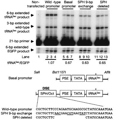
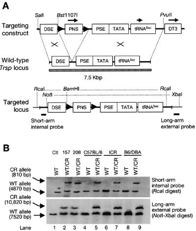
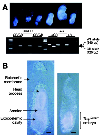
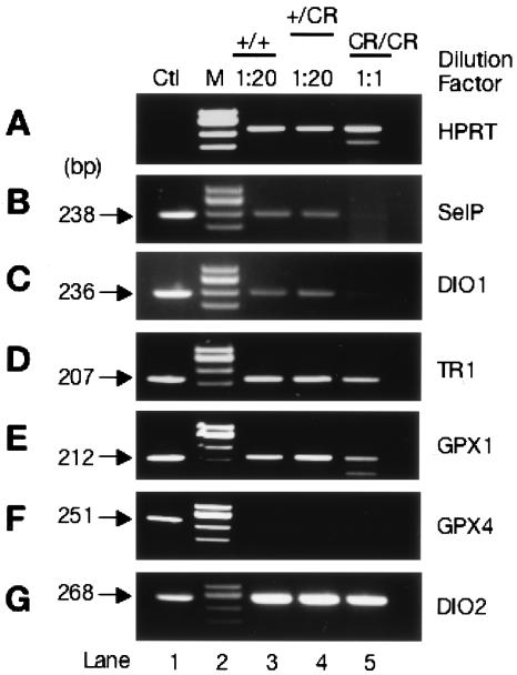
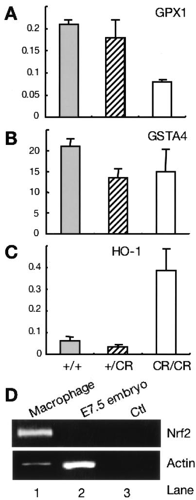
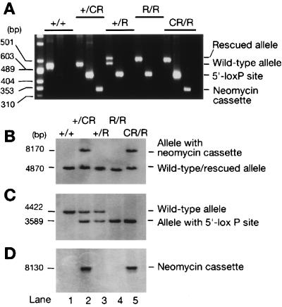
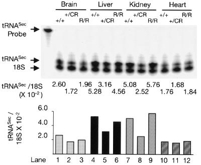
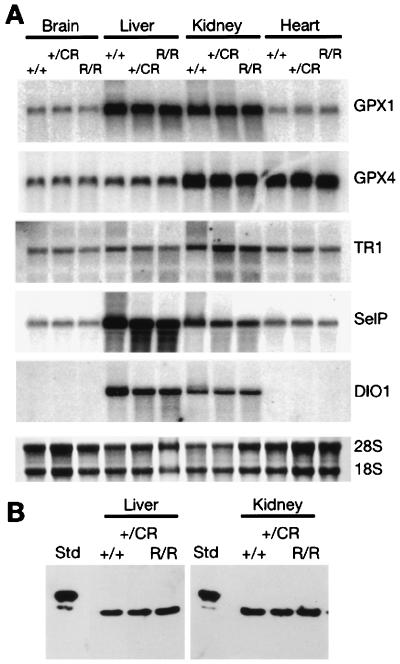
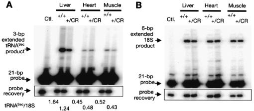
Appropriate expression of the selenocysteine tRNA (tRNA(Sec)) gene is necessary for the production of an entire family of selenoprotein enzymes. This study investigates the consequence of disrupting an upstream enhancer region of the mouse tRNA(Sec) gene (Trsp) known as the distal sequence element (DSE) by use of a conditional repair gene targeting strategy, in which a 3.2-kb insertion was introduced into the promoter of the gene. In the absence of DSE activity, homozygous mice failed to develop in utero beyond embryonic day 7.5 and had severely decreased levels of selenoprotein transcript. Cre-mediated removal of the selection cassette recovered DSE regulation of Trsp, restoring wild-type levels of tRNA(Sec) expression and allowing the generation of viable rescued mice. Further analysis of targeted heterozygous adult mice revealed that the enhancer activity of the DSE is tissue dependent since, in contrast to liver, heart does not require the DSE for normal expression of Trsp. Similarly, in mouse cell lines we showed that the DSE functions as a cell-line-specific inducible element of tRNA(Sec). Together, our data demonstrate that the DSE is a tissue-dependent regulatory element of tRNA(Sec) expression and that its activity is vital for sufficient tRNA(Sec) production during mouse embryogenesis.


Figures

FIG. 1.
FIG. 1.
Analysis of tRNASec transcriptional regulation by the DSE and SPH motif in mouse NIH 3T3 cells. NIH 3T3 cells were transfected with EGFP reporter construct and Trsp reporter plasmid that has a wild-type promoter (Wild-type promoter), lacks the DSE region (basal promoter), harbors a 9-bp exchange in the SPH element (SPH 9-bp exchange), or has a 15-bp deletion within the SPH element (SPH deleted). The octamer motif, positioned immediately downstream of the SPH element, is shown in italics. The endogenous tRNASec molecule yields a 3-bp product by extension from a specific 21-bp primer, whereas the tRNASec molecule arising from the transfected constructs generates a 6-bp extension product due to a T-to-C mutation in the tRNASec coding sequence, indicated by asterisks. Primer extension of the EGFP transcript generates a 6-bp extended product. The structure of the reporter constructs and the sequence of the wild-type or mutant SPH elements are shown.
FIG. 2.
FIG. 2.
Generation of Trsp conditional repair mice. (A) Simplified targeting strategy, not drawn to scale, for homologous recombination in ES cells showing the targeting vector, the wild-type Trsp gene, and the targeted allele. Open boxes denote the functional elements described in the text. Overhead arrows depict the orientation of transcriptional units. The PNS cassette is shown flanked by loxP elements, designated by a solid arrowhead. Black bars show the genomic fragments used as probes for Southern blot hybridization analyses presented in panel B. Only relevant restriction sites are shown. (B) Demonstration of correct homologous recombination in two independent ES clones and genotype analysis of mouse strains carrying the targeted insertion by Southern blot hybridization. DNA digested with RcaI was probed by using a short-arm internal probe (upper panel), and DNA digested with NotI and XbaI was probed by using a long-arm external probe (lower panel). The gels were loaded as follows: parental ES cell line (lane 1; Ctl), homologous recombinant clones number 157 and 208 (lanes 2 and 3, respectively), DNA from adult wild type (WT) and germ line heterozygotes (WT/CR) on a C57BL/6 background (lanes 4 and 5), ICR background (lanes 6 and 7), and a mixed C57BL/6 DBA background (lanes 8 and 9).
FIG. 3.
FIG. 3.
Analysis of embryos generated from a Trsp+/CR intercross. (A) E7.5 embryos were fixed briefly in formaldehyde before removal of maternal and extra-embryonic tissues. The embryos were photographed, and genomic DNA was extracted. PCR genotyping was performed with the primer pair SeCKDCRFP1 and SeCKDCRP1, yielding a 540-bp product from the wild-type allele and SeCKDCRFP1 and KDRP1, generating a 420-bp product from the CR targeted allele. (B) High-resolution photograph of wild-type (left) and homozygous mutant E7.5 embryo (right). Bar, 100 μm.
FIG. 4.
FIG. 4.
RT-PCR analysis of selenoprotein transcript levels in Trsp+/+, Trsp+/CR, and TrspCR/CR embryos. E7.5 embryos were dissected from maternal and extra-embryonic tissues, and total RNA was extracted. RNA from three embryos of each genotype was pooled and reverse transcribed. The HPRT PCR product was used as a loading control to equalize RNA input levels in each of the RTPCR reactions. A 20-fold-greater quantity of total RNA from TrspCR/CR embryos was required to equalize that of Trsp+/+and Trsp+/CR embryos. Primer pairs specific to each of the selenoprotein species were used to amplify the RNA message of the indicated proteins. cDNA for each of the selenoproteins was used as a positive control (Ctl) in the PCRs. DNA size markers (M) were φX174 DNA digested with HaeIII. The molecular size (in base pairs) of each PCR product is presented on the left-hand side of the figure.
FIG. 5.
FIG. 5.
Expression of GPX1, GSTA4, HO-1, and Nrf2 in Trsp+/+, Trsp+/CR, and TrspCR/CR embryos. The expression profiles of GPX1 (A), GSTA4 (B), and HO-1 (C) were analyzed by quantitative real-time RT-PCR with rRNA as the normalization control. (D) RT-PCR was used to examine the expression of Nrf2 and β-actin in E7.5 wild-type mice (lane 2), with peritoneal macrophage serving as the positive control (lane 1) and water as a negative control (lane 3).
FIG. 6.
FIG. 6.
Generation of Trsp+/R, TrspR/R, and TrspR/CR mice. Mice carrying the AyuI-Cre transgene were crossed with Trsp+/CR mice to generate heterozygous Trsp+/R animals. Subsequently, heterozygous rescue mice were intercrossed with Trsp+/R and Trsp+/CR mice to generate homozygous TrspR/R and TrspR/CR mice, respectively. (A) Genomic DNA was extracted from mouse tail samples and amplified by PCR. The primers used to detect the wild-type allele were SeCKDCRFP1 and SeCKDCRP1, and the 5′-loxP site SeCKDCRFP1 and KDRP1, and for the neomycin phosphotransferase gene were Neo5-Prime and Neo3-Prime as described in Table 1. On the right-hand side of the panel the identity of each PCR product is indicated. After Cre-mediated recombination of the CR allele a single loxP element (50 bp, including flanking sequences) remains, positioned between the DSE and PSE. Therefore, the PCR product arising from the rescued allele is distinguishable from that of the wild-type allele due to its lower mobility. For Southern blot analysis mice that had been selected by PCR were sacrificed, and liver genomic DNA was recovered. (B and C) Genomic DNA was digested with RcaI (B) or digested simultaneously with RcaI and BamHI (a single site exits external to the loxP element) (C), and the transfer membrane was probed by using the SspI-SwaI internal probe. (D) RcaI was used to digest the DNA, and a XbaI-PstI fragment from the neomycin phosphotransferase gene was used as a probe.
FIG. 7.
FIG. 7.
Quantification of tRNASec levels in adult Trsp+/+, Trsp+/CR, and TrspR/R mice by RNase protection assay. Antisense RNA to tRNASec (240 bp) and the 18S ribosomal subunit (130 bp) were prepared and hybridized overnight to 2.5 μg of total RNA from brains, livers, kidneys, and hearts. RNase digestions were performed by using an RNase A-RNase T1 mix, and the protected fragments were resolved on a 10% denaturing polyacrylamide gel. The migratory position of the protected fragment for tRNASec (91 bp) and the 18S subunit (80 bp) are indicated on the left-hand side of the figure. The relative values of the transcript signals (tRNASec/18S) were obtained by densitometric analyses. The values presented have been adjusted to account for a 25-fold-greater specific activity of the tRNASec probe relative to the 18S ribosomal probe.
FIG. 8.
FIG. 8.
Determination of selenoprotein transcript levels and GPX1 protein levels in Trsp+/+, Trsp+/CR, and TrspR/R mice. (A) Total RNA was extracted from the brains, livers, kidneys, and hearts of the indicated mouse strains, and RNA blot analyses were performed with cDNA for the selenoprotein species listed on the right-hand side of the panel. The 18S ribosomal unit, stained with methylene blue, was used as a loading control. (B) Cytosolic extract from the livers and kidneys was immunoblotted with anti-GPX1 antibody. Recombinant polyhistidine-tagged GPX1 was used as standard (Std). Sodium dodecyl sulfate-polyacrylamide gel electrophoresis and Coomassie blue staining of cytosolic extracts were performed separately to confirm equivalent loading (data not shown).
FIG. 9.
FIG. 9.
Primer extension analysis of tRNASec in Trsp+/+ and Trsp+/CR mice. Total RNA was isolated from livers, hearts, and muscles. (A and B) A radiolabeled 21-bp tRNASec specific primer was used to generate a 3-bp extension product (A), whereas a 21-bp 18S specific primer was used to yield a 6-bp elongated product (B). Primer and products were resolved on a 20% denaturing polyacrylamide gel. In the control lane (Ctl), no RNA had been added.
FIG. 10.
FIG. 10.
The expression of tRNASec correlates with DSE binding activity. (A) Total RNA extracted from the various mouse cell lines was analyzed for tRNASec levels by primer extension. The tRNASec primer yields a 3-bp extension product, whereas the 18S primer generates a 6-bp extension product. (B) Binding assay on wild-type and mutant mouse Trsp fragments encompassing the DSE with nuclear protein extracts from the indicated mouse cell lines. (C) Total RNA from the adherent (A), mixed adherent-suspension (M), and suspension (S) mouse cell lines was examined for Staf transcript levels by RNA blot analysis. Methylene blue staining for the 18S ribosomal subunit served as a loading control.

Similar articles

Cited by

References

    1. Adachi, K., H. Saito, T. Tanaka, and T. Oka. 1998. Molecular cloning and characterization of the murine staf cDNA encoding a transcription activating factor for the selenocysteine tRNA gene in mouse mammary gland. J. Biol. Chem. 273:8598-8606. - PubMed
    1. Bianco, A. C., D. Salvatore, B. Gereben, M. J. Berry, and P. R. Larsen. 2002. Biochemistry, cellular and molecular biology, and physiological roles of the iodothyronine selenodeiodinases. Endocrinol. Rev. 23:38-89. - PubMed
    1. Bosl, M. R., M. F. Seldin, S. Nishimura, and M. Taketo. 1995. Cloning, structural analysis and mapping of the mouse selenocysteine tRNA([Ser]Sec) gene (Trsp). Mol. Gen. Genet. 248:247-252. - PubMed
    1. Bosl, M. R., K. Takaku, M. Oshima, S. Nishimura, and M. M. Taketo. 1997. Early embryonic lethality caused by targeted disruption of the mouse selenocysteine tRNA gene (Trsp). Proc. Natl. Acad. Sci. USA 94:5531-5534. - PMC - PubMed
    1. Carbon, P., and A. Krol. 1991. Transcription of the Xenopus laevis selenocysteine tRNA(Ser)Sec gene: a system that combines an internal B box and upstream elements also found in U6 snRNA genes. EMBO J. 10:599-606. - PMC - PubMed

Publication types

MeSH terms

LinkOut - more resources