. 2005 May;25(9):3752-62.
doi: 10.1128/MCB.25.9.3752-3762.2005.

Cyclins D2 and D1 are essential for postnatal pancreatic beta-cell growth

Affiliations

Cyclins D2 and D1 are essential for postnatal pancreatic beta-cell growth

Jake A Kushner et al. Mol Cell Biol. 2005 May.

Abstract

Regulation of adult beta-cell mass in pancreatic islets is essential to preserve sufficient insulin secretion in order to appropriately regulate glucose homeostasis. In many tissues mitogens influence development by stimulating D-type cyclins (D1, D2, or D3) and activating cyclin-dependent kinases (CDK4 or CDK6), which results in progression through the G(1) phase of the cell cycle. Here we show that cyclins D2 and D1 are essential for normal postnatal islet growth. In adult murine islets basal cyclin D2 mRNA expression was easily detected, while cyclin D1 was expressed at lower levels and cyclin D3 was nearly undetectable. Prenatal islet development occurred normally in cyclin D2(-/-) or cyclin D1(+/-) D2(-/-) mice. However, beta-cell proliferation, adult mass, and glucose tolerance were decreased in adult cyclin D2(-/-) mice, causing glucose intolerance that progressed to diabetes by 12 months of age. Although cyclin D1(+/-) mice never developed diabetes, life-threatening diabetes developed in 3-month-old cyclin D1(-/+) D2(-/-) mice as beta-cell mass decreased after birth. Thus, cyclins D2 and D1 were essential for beta-cell expansion in adult mice. Strategies to tightly regulate D-type cyclin activity in beta cells could prevent or cure diabetes.


Figures

FIG. 1.
FIG. 1.
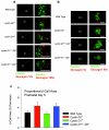
D-type cyclin content analysis in mouse islets. (a) RT-PCR analysis of D-type cyclin RNA content in wild-type male mouse islets expressed as fraction of cyclin D2 expression. Results are expressed as means ± SEM for six samples per group. (b) Representative immunohistochemistry of wild-type male mouse pancreas section stained with guinea pig anti-insulin (green) and mouse anti-cyclin D2 (red) antisera. Scale bar: 100 μm.
FIG. 2.
FIG. 2.
Neonatal islet histology and morphometric analysis. (a) Representative islet histology of pancreas sections from 5-day-old wild-type, cyclin D2/, cyclin D1+/−, cyclin D1+/− D2/, and cyclin D1/ D2+/− mice. Immunostaining with antibodies against insulin (green) and glucagon (red) at ×10 (left panels) and at ×100 (right panels). Scale bars: 100 μm. (b) Sixteen-day-old wild-type, cyclin D2/, cyclin D1+/− D2/, and cyclin D1/ D2/ mice. Immunostaining with antibodies against insulin (green) and glucagon (red) at ×5 (left panels) and at ×40 (right panels). Scale bars: 100 μm. (c) Mean proportional cross-sectional β-cell area (reported as percentage of total pancreas area) of wild-type, cyclin D2/, cyclin D1+/−, and cyclin D1+/− D2/ mice at postnatal day 5.
FIG. 3.
FIG. 3.
Metabolic testing of male wild-type and cyclin D2/ mice. (a to c) Glucose tolerance tests of male wild-type and cyclin D2/ mice. Serum blood glucose measurements expressed as mg/dl of 3-month-old mice (a) and 9- to 12-month-old mice (b) after intraperitoneal injection of 2 g d-glucose/kg body weight. *, P < 0.05, cyclin D2/ versus wild-type; **, P < 0.01, cyclin D2/ versus wild type. (c to d) Insulin tolerance tests of male wild-type and cyclin D2−/− mice. (c) Serum blood glucose measurements expressed as means ± SEM of percentages of initial blood glucose values of 3-month-old (c) or 9- to 12-month-old (d) male wild-type and cyclin D2/ mice after intraperitoneal injection of 1.5 U/kg human regular insulin. Results represent at least 10 mice per group, except wild-type mice at 9 to 12 months (n = 5).
FIG. 4.
FIG. 4.
Metabolic testing of cyclin D1+/− D2+/− intercross mice. (a) Glucose tolerance tests of male wild-type, cyclin D2/, cyclin D1+/−, and cyclin D1+/− D2/ mice at 3 months of age. Serum blood glucose measurements are expressed as mg/dl after intraperitoneal injection of 2 g d-glucose/kg body weight. (b) Insulin tolerance tests of male wild-type, cyclin D2/, and cyclin D1+/− mice at 3 months of age. Serum blood glucose measurements expressed as means ± SEM of percentages of initial blood glucose values after intraperitoneal injection of 1.5 U/kg human regular insulin. Results represent at least 10 mice per group, except cyclin D1+/− mice (n = 5).
FIG.5.
FIG.5.
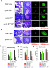
Islet histology and morphometric analysis of adult wild-type, cyclin D2/, cyclin D1+/−, and cyclin D1+/− D2/ mice. (a) Representative islet histology from pancreas sections from 3-month-old male mice. Hematoxylin and eosin (H&E) staining at ×5 (furthest left panels) and at ×40 (second from left panels). Immunostaining with antibodies against insulin (green) and glucagon (red) at ×5 (second from right panels) and at ×40 (furthest right panels). Scale bars: 100 μm. (b) Representative islet histology from pancreas sections from 9- to 12-month-old male mice. Hematoxylin and eosin (H&E) staining at ×5 (furthest left panels) and at ×40 (second from left panels). Immunostaining with antibodies against insulin (green) and glucagon (red) at ×5 (second from right panels) and at ×40 (furthest right panels). Scale bars: 100 μm. (c to e) Islet morphometric studies of wild-type, cyclin D2/, cyclin D1+/−, and cyclin D1+/− D2/ mice at 3 months and wild-type and cyclin D2/ mice at 9 to 12 months, with at least 4 to 5 mice per group analyzed. (c) Mean cross-sectional β-cell area, reported as percentage of total pancreas area. **, P < 0.01, cyclin D2/ versus wild-type. (d) Islet size calculated by mean cross-sectional area of multicelled islets reported as microns ×103/islet. **, P < 0.01, cyclin D2/ versus wild type. (e) Islet density calculated reported as islets per mm2. *, P < 0.05, cyclin D2/ versus wild type.
FIG. 6.
FIG. 6.
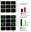
Neonatal and adult islet proliferation analysis of wild-type, cyclin D2/, cyclin D1+/−, and cyclin D1+/− D2+/− mice. (a) Representative histologic islet proliferation analysis of pancreas sections from 16-day-old male mice stained with insulin (furthest left panels), BrdU (with islets outlined) (middle panels), and insulin/BrdU images merged (right panels). Proliferating β cells noted by white triangles. Images photographed at ×40, scale bars: 100 μm. (b) Percent BrdU incorporation in β cells measured at 16 days of life per genotype. **, P < 0.01, cyclin D1+/− D2/ versus cyclin D2/ or wild type. Results are expressed as the means ± SEM fraction of BrdU/insulin copositive β cells compared to total β cells. (c) BrdU incorporation in β cells at 3 months of age per genotype. **, P < 0.01, cyclin D2/ versus wild type. Results are expressed as the means ± SEM fraction of BrdU/insulin-copositive β cells compared to total β cells. (d) Representative histologic islet proliferation analysis of pancreas sections from 3-month-old male mice stained with insulin (furthest left panels), BrdU (with islets outlined) (middle panels), and insulin/BrdU images merged (right panels).
FIG. 7.
FIG. 7.
D-type cyclin mRNA expression in pancreatic islets from cyclin D2/ mice and controls. TaqMan RT-PCR of cyclin D1, D2, and D3 with islet mRNA from male cyclin D2/ mice (n = 4) and wild-type controls (n = 4) at 6 weeks of age. Data are normalized to cyclophilin gene expression and expressed as means ± SEM.

References

    1. Bell, G. I., and K. S. Polonsky. 2001. Diabetes mellitus and genetically programmed defects in beta-cell function. Nature 414:788-791. - PubMed
    1. Bonner-Weir, S. 2001. Beta-cell turnover: its assessment and implications. Diabetes 50(Suppl. 1):S20-S24. - PubMed
    1. Bonner-Weir, S., E. Toschi, A. Inada, P. Reitz, S. Y. Fonseca, T. Aye, and A. Sharma. 2004. The pancreatic ductal epithelium serves as a potential pool of progenitor cells. Pediatr. Diabetes 5(Suppl. 2):16-22. - PubMed
    1. Butler, A. E., J. Janson, S. Bonner-Weir, R. Ritzel, R. A. Rizza, and P. C. Butler. 2003. Beta-cell deficit and increased beta-cell apoptosis in humans with type 2 diabetes. Diabetes 52:102-110. - PubMed
    1. Ciemerych, M. A., A. M. Kenney, E. Sicinska, I. Kalaszczynska, R. T. Bronson, D. H. Rowitch, H. Gardner, and P. Sicinski. 2002. Development of mice expressing a single D-type cyclin. Genes Dev. 16:3277-3289. - PMC - PubMed

Publication types

MeSH terms