. 2005 May;25(9):3814-30.
doi: 10.1128/MCB.25.9.3814-3830.2005.

The human stress-activated protein kin17 belongs to the multiprotein DNA replication complex and associates in vivo with mammalian replication origins

Affiliations

The human stress-activated protein kin17 belongs to the multiprotein DNA replication complex and associates in vivo with mammalian replication origins

Laurent Miccoli et al. Mol Cell Biol. 2005 May.

Abstract

The human stress-activated protein kin17 accumulates in the nuclei of proliferating cells with predominant colocalization with sites of active DNA replication. The distribution of kin17 protein is in equilibrium between chromatin-DNA and the nuclear matrix. An increased association with nonchromatin nuclear structure is observed in S-phase cells. We demonstrated here that kin17 protein strongly associates in vivo with DNA fragments containing replication origins in both human HeLa and monkey CV-1 cells. This association was 10-fold higher than that observed with nonorigin control DNA fragments in exponentially growing cells. In addition, the association of kin17 protein to DNA fragments containing replication origins was also analyzed as a function of the cell cycle. High binding of kin17 protein was found at the G(1)/S border and throughout the S phase and was negligible in both G(0) and M phases. Specific monoclonal antibodies against kin17 protein induced a threefold inhibition of in vitro DNA replication of a plasmid containing a minimal replication origin that could be partially restored by the addition of recombinant kin17 protein. Immunoelectron microscopy confirmed the colocalization of kin17 protein with replication proteins like RPA, PCNA, and DNA polymerase alpha. A two-step chromatographic fractionation of nuclear extracts from HeLa cells revealed that kin17 protein localized in vivo in distinct protein complexes of high molecular weight. We found that kin17 protein purified within an approximately 600-kDa protein complex able to support in vitro DNA replication by means of two different biochemical methods designed to isolate replication complexes. In addition, the reduced in vitro DNA replication activity of the multiprotein replication complex after immunodepletion for kin17 protein highlighted for a direct role in DNA replication at the origins.


Figures
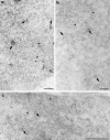
FIG. 1.
FIG. 1.
Ultrastructural colocalization of kin17 protein with DNA replication proteins. Ultrathin sections were prepared from proliferating RKO cells for immunoelectron microscopy as indicated in Materials and Methods. kin17 protein was identified with a mixture of two monoclonal antibodies (purified K36 and K58 antibodies). Large gold particles (10 nm) indicate the incorporation of RPA (a), PCNA (b), or DNA polymerase α (c), and small gold particles (5 nm) reveal kin17 protein labeling. Arrows point to colocalization of kin17 and RPA, kin17 and PCNA, or kin17 and DNA polymerase α; arrowheads point to kin17 alone. Representative microphotographs are shown. Scale bars, 0.5 μm.
FIG. 2.
FIG. 2.
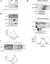
kin17 protein is a component of high-molecular-weight nuclear protein complexes. (A) Immunoblot analysis and chromatographic profiles of HeLa S3 NE fractionated on a Superose 6 GF column in the absence [BET(−)] and in the presence [BET(+)] of BET. (B) Immunoblot analysis of HeLa S3 NE fractionated on a Mono Q column and step eluted in 0.2 M KCl, 0.3 M KCl, and 1 M KCl. (C) Mono Q-eluted FT, 0.3 M KCl, and 1 M KCl fractions no. 3 were then applied to a Superose 6 GF column in equilibration buffer containing 0.1 M KCl, 0.3 M KCl, and 1 M KCl, respectively. An equal volume of each collected fraction was loaded onto a 10% SDS-polyacrylamide gel, followed by Western blotting with anti-kin17, anti-RPA, and anti-PCNA antibodies. Protein molecular size standards were analyzed under the same conditions. The molecular mass of protein complexes was determined from the calibration curve of the molecular masses of protein standards.
FIG. 3.
FIG. 3.
kin17 protein is a component of the MRC in human HeLa cells. (A) Immunoblot analysis for the presence of kin17 protein in fractions from purification of the human MRC. Extracted nuclear proteins were centrifuged onto a 2 M sucrose cushion and divided into an interphase fraction consisting of high-molecular-weight protein complexes (including the MRC referred to as fraction HSP-4) and a supernatant fraction consisting of soluble proteins (fraction HS-4). Ten fractions were collected. (B) Mono Q elution profile. Fractions 9 and 10 were pooled (HSP-4) and loaded onto a Mono Q column. Immunoblot analysis and ATPase and polymerase activity profiles of Mono Q fractions are shown. (C) GFelution profile of the HSP-4 fraction. Seventy-five micrograms of the HSP-4 fraction was loaded onto a Superose 6 column. Immunoblot analysis for the presence of kin17 protein and ATPase (filled circles) and polymerase activity (open squares) profiles of GF fractions are shown. (His)6kin17 purified protein was used as a positive control. (D) GF elution profile of the HSP-4 fraction pretreated with both DNase and RNase. Seventy-five micrograms of the HSP-4 fraction pretreated with both DNase and RNase was loaded onto a Superose 6 column. Immunoblot analysis of kin17 protein, RPA 70, and PCNA, and polymerase activity with both DNase and RNase (filled squares) and without DNase and RNase (open squares) of GF fractions are shown.
FIG.4.
FIG.4.
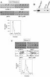
kin17 protein copurifies with an RC complex from S-phase HeLa cells. (A) GF elution profile. Dot blot analysis of GF fractions of the 0.2 M NaCl NE from HeLa cells was performed with antibodies against polymerase α (pol α), RFC, cyclin A, and kin17. Polymerase (Pol) activity was assayed in these fractions. L, loading. (B) Western blot analysis of GF peak fractions isolated from asynchronous (AS) and S-phase HeLa cells with anti-kin17 antibodies. (His)6-kin17 recombinant protein was used as a positive control (C) Mono S elution profile. Fractions from the GF containing DNA polymerase activity and proteins of interest were pooled and loaded onto a Mono S column, which was eluted with a linear NaCl gradient. The Mono S fractions were tested for polymerase activity and subjected to immunoblot analysis with antibodies against polymerase α, RFC, cyclin A, and kin17. (D) Heparin-Sepharose elution profile. Active fractions from the Mono S column were pooled and loaded onto a heparin column. Immunoblot analysis and polymerase activity profile of heparin fractions are shown. (E) Mono Q elution profile. Active fractions from the heparin-Sepharose column were pooled and loaded onto a Mono Q column. Immunoblot analysis and polymerase activity profile of Mono Q fractions are shown. All chromatographic steps were performed at 4°C as described in Materials and Methods. (F) Western blot analysis of the GF, Mono S, heparin-Sepharose, and Mono Q peak fractions that contained the RC complex with antibodies against RFC and kin17 protein. (G) Twenty micrograms of fraction 6 of GF were resolved through 4 to 15% native polyacrylamide gel electrophoresis and electrophoretically transferred to nitrocellulose. The native Western blots were probed with antibodies against kin17 protein, RPA, and DNA polymerase α. (H) In vivo cross-linking of replicative complexes in S-phase HeLa cells. Free proteins and DNA-protein complexes were extracted as described in Materials and Methods. Western blot analysis was performed on DNA-protein complexes and in fractions that contained free proteins using antibodies against kin17, RPA, cyclin A, and PCNA proteins.
FIG.4.
FIG.4.
kin17 protein copurifies with an RC complex from S-phase HeLa cells. (A) GF elution profile. Dot blot analysis of GF fractions of the 0.2 M NaCl NE from HeLa cells was performed with antibodies against polymerase α (pol α), RFC, cyclin A, and kin17. Polymerase (Pol) activity was assayed in these fractions. L, loading. (B) Western blot analysis of GF peak fractions isolated from asynchronous (AS) and S-phase HeLa cells with anti-kin17 antibodies. (His)6-kin17 recombinant protein was used as a positive control (C) Mono S elution profile. Fractions from the GF containing DNA polymerase activity and proteins of interest were pooled and loaded onto a Mono S column, which was eluted with a linear NaCl gradient. The Mono S fractions were tested for polymerase activity and subjected to immunoblot analysis with antibodies against polymerase α, RFC, cyclin A, and kin17. (D) Heparin-Sepharose elution profile. Active fractions from the Mono S column were pooled and loaded onto a heparin column. Immunoblot analysis and polymerase activity profile of heparin fractions are shown. (E) Mono Q elution profile. Active fractions from the heparin-Sepharose column were pooled and loaded onto a Mono Q column. Immunoblot analysis and polymerase activity profile of Mono Q fractions are shown. All chromatographic steps were performed at 4°C as described in Materials and Methods. (F) Western blot analysis of the GF, Mono S, heparin-Sepharose, and Mono Q peak fractions that contained the RC complex with antibodies against RFC and kin17 protein. (G) Twenty micrograms of fraction 6 of GF were resolved through 4 to 15% native polyacrylamide gel electrophoresis and electrophoretically transferred to nitrocellulose. The native Western blots were probed with antibodies against kin17 protein, RPA, and DNA polymerase α. (H) In vivo cross-linking of replicative complexes in S-phase HeLa cells. Free proteins and DNA-protein complexes were extracted as described in Materials and Methods. Western blot analysis was performed on DNA-protein complexes and in fractions that contained free proteins using antibodies against kin17, RPA, cyclin A, and PCNA proteins.
FIG.5.
FIG.5.
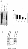
Immunodepletion of kin17 protein inhibits in vitro replication of SV40 origin- and large T antigen-dependent DNA replication. The DNA replication activity of immunodepleted and mock-immunodepleted HSP-4 fraction for the kin17 protein was assessed by the system of Li and Kelly (36). Immunodepletion of kin17 protein was performed on HSP-4 fractions and were incubated with plasmid pUC.HSO, which contains a functional SV40 origin of replication, in replication buffer at 37°C for 4 h. At the end of the incubation period, an aliquot was removed from each reaction mixture and the incorporation of labeled nucleotides into DNA was assessed by precipitation with 10% trichloroacetic acid and scintillation counting. The remainder of each reaction mixture was run on a 1% agarose gel and visualized by PhosphorImager. Results are expressed as a percentage of the control (mock immunodepleted; 100%). (A) Autoradiography of a DNA replication assay with immunodepleted extracts. Lane 1, HSP-4 Tag(+), positive control 100%, mock-immunodepleted extract; lane 2, HSP-4 Tag(−), negative control without large T antigen; lane 3, Id-HSP-4 Tag(+), kin17-immunodepleted HSP-4 extract. (B) Quantification of the inhibition of DNA replication assays by the immunodepletion of HSP-4 extracts by anti-kin17 (lane Id-kin17), anti-isotype control (lane Id-isotype), and anti-PCNA antibodies (lane Id-PCNA). Results are expressed as a percentage of the control (HSP-4 fraction; 100%). (C) Western blot analysis of PCNA- and kin17-immunodepleted HSP-4 fraction. IP anti-kin17, immunoblot analysis of kin17 protein, RPA 70, and PCNA after immunoprecipitation of HSP-4 fraction with anti-kin17 antibodies; IP anti-PCNA, immunoblot analysis of PCNA, kin17 protein, and RPA 70 after immunoprecipitation of HSP-4 fraction with anti-PCNA antibodies; IP anti-isotype, immunoblot analysis of kin17 protein, RPA 70, and PCNA after immunoprecipitation of HSP-4 fraction anti-isotype antibodies. HC, heavy-chain immunoglobulin.
FIG.6.
FIG.6.
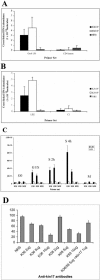
Direct involvement of kin17 protein in DNA replication. (A, B) kin17 protein associates in vivo with mammalian DNA replication origins. Quantification of DNA abundance in origin-containing and non-origin-containing sequences by real-time PCR is shown. In vivo association of kin17 protein with monkey and human origins of replication was analyzed by a chromatin immunoprecipitation (IP) assay and quantitative PCR analysis. (A) Total normalized cross-linked molecules detected by real-time PCR with primer sets ors8 150 and CD4 intron from exponentially growing CV-1 cells cross-linked and immunoprecipitated with monoclonal antibodies K36 and K58 against kin17 protein and with NRS. (B) Total normalized cross-linked molecules detected by real-time PCR with primer sets LB2 and C1 from exponentially growing HeLa cells cross-linked and immunoprecipitated with monoclonal antibodies K36 and K58 against the kin17 protein and with NRS. (C) Total normalized cross-linked molecules detected by real-time PCR with primer sets LB2 and C1 from HeLa cells in different phases of the cell cycle cross-linked and immunoprecipitated with monoclonal antibodies K36 and K58 against kin17 protein and with NRS. (D) Anti-kin17 antibodies inhibit in vitro DNA replication of p186. Effects of addition of increasing amounts (2, 5, or 10 μg) of anti-kin17 antibodies (K36 and K58) and of recombinant human kin17 protein (1 μg) to the in vitro reaction mixture on p186 replication, by comparison to NRS, are shown.

Similar articles

Cited by

References

    1. Alvarez, D., M. Callejo, R. Shoucri, L. Boyer, G. B. Price, and M. Zannis-Hadjopoulos. 2003. Analysis of the cruciform binding activity of recombinant 14-3-3ζ-MBP fusion protein, its heterodimerization profile with endogenous 14-3-3 isoforms, and effect on mammalian DNA replication in vitro. Biochemistry 42:7205-7215. - PubMed
    1. Angulo, J. F., E. Rouer, A. Mazin, M. G. Mattei, A. Tissier, P. Horellou, R. Benarous, and R. Devoret. 1991. Identification and expression of the cDNA of KIN17, a zinc-finger gene located on mouse chromosome 2, encoding a new DNA-binding protein. Nucleic Acids Res. 19:5117-5123. - PMC - PubMed
    1. Araujo, F. D., J. D. Knox, S. Ramchandani, R. Pelletier, P. Bigey, G. Price, M. Szyf, and M. Zannis-Hadjopoulos. 1999. Identification of initiation sites for DNA replication in the human dnmt1 (DNA-methyltransferase) locus. J. Biol. Chem. 274:9335-9341. - PubMed
    1. Batty, D. P., and R. D. Wood. 2000. Damage recognition in nucleotide excision repair of DNA. Gene 241:193-204. - PubMed
    1. Berezney, R., M. J. Mortillaro, H. Ma, X. Wei, and J. Samarabandu. 1995. The nuclear matrix: a structural milieu for genomic function. Int. Rev. Cytol. 162A:1-65. - PubMed

Publication types

MeSH terms

LinkOut - more resources