. 2005 May;25(9):3831-41.
doi: 10.1128/MCB.25.9.3831-3841.2005.

Dok-R mediates attenuation of epidermal growth factor-dependent mitogen-activated protein kinase and Akt activation through processive recruitment of c-Src and Csk

Affiliations

Dok-R mediates attenuation of epidermal growth factor-dependent mitogen-activated protein kinase and Akt activation through processive recruitment of c-Src and Csk

Paul Van Slyke et al. Mol Cell Biol. 2005 May.

Abstract

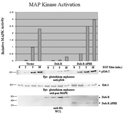
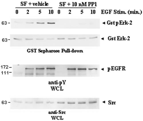
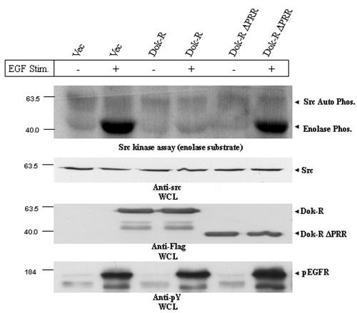
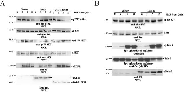
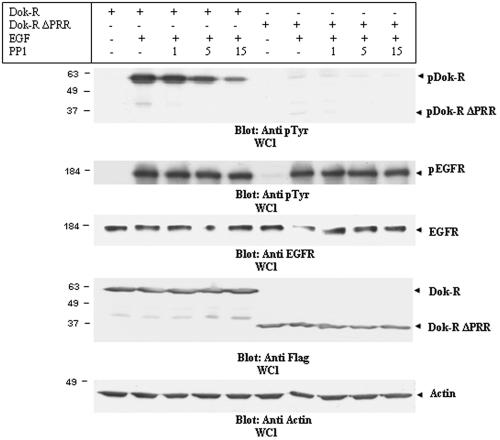
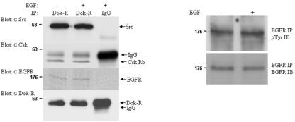
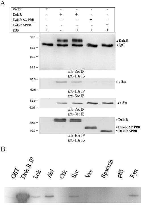
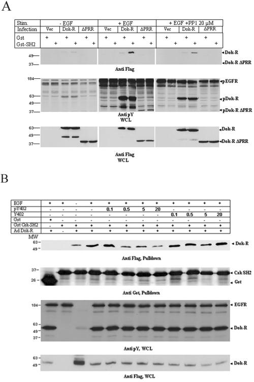
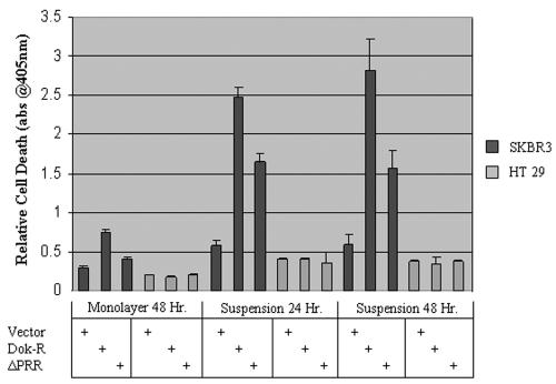
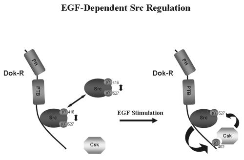
Dok-R has previously been shown to associate with the epidermal growth factor receptor (EGFR) and become tyrosine phosphorylated in response to EGF stimulation. The recruitment of Dok-R to the EGFR, which is mediated through its phosphotyrosine binding (PTB) domain, results in attenuation of mitogen-activated protein kinase (MAPK) activation. Dok-R's ability to attenuate EGF-driven MAPK activation is independent of its ability to recruit rasGAP, a known attenuator of MAPK activity, suggesting an alternate Dok-R-mediated pathway. Herein, we have determined the structural determinants within Dok-R that are required for its ability to attenuate EGF signaling and to associate with c-Src and with the Src family kinase (SFK)-inhibitory kinase, Csk. We demonstrate that Dok-R associates constitutively with c-Src through an SH3-dependent interaction and that this association is essential to Dok-R's ability to attenuate c-Src activity and diminish MAPK and Akt/PKB activity. We further illustrate that EGF-dependent phosphorylation of Dok-R requires SFK activity and, more specifically, that SFK-dependent phosphorylation of tyrosine 402 on Dok-R facilitates the inducible recruitment of Csk. We propose that recruitment of Csk to Dok-R serves to bring Csk to c-Src and down-regulate its activity, resulting in a concomitant attenuation of MAPK and Akt/PKB activity. Furthermore, we demonstrate that Dok-R can abrogate c-Src's ability to protect the breast cancer cell line SKBR3 from anoikis and that an association with c-Src and Csk is required for this activity. Collectively these results demonstrate that Dok-R acts as an EGFR-recruited scaffolding molecule that processively assembles c-Src and Csk to attenuate signaling from the EGFR.


Figures

FIG. 1.
FIG. 1.
The carboxy-terminal region of Dok-R is required for suppression of EGF-dependent MAPK activation. Cos1 cells were transiently transfected with GST-Erk-2 and either vector Dok-R or Dok-R ΔPRR. Serum-starved Cos1 cells were left unstimulated (lane 0) or stimulated (Stim) with 100 ng/ml of EGF for the indicated times (2, 5, or 10 min). Cleared lysates were prepared, and glutathione Sepharose was used to specifically purify the exogenous GST-Erk-2. Immunoblot analysis with phospho-specific MAPK, pan-MAPK (GST-pull-downs) as well as anti-HA (WCL) was performed. The graph represents relative MAPK activation determined via anti-phosho-MAPK immunoblot analysis when normalized to pan-Erk-2 levels using densitometry (Molecular Dynamics and ImageQuant 5.0). Results are representative of four independent experiments and serve to demonstrate that the PRR of Dok-R is necessary to attenuate EGF-dependent MAPK activation.
FIG. 2.
FIG. 2.
SFK activity is required for full activation of EGF-dependent MAPK activity. GST-Erk-2-transfected Cos1 cells were serum starved (serum free [SF]) for 16 h prior to being treated with SFK inhibitor PP1 (10 μM) or vehicle (DMSO) for 2 h. Cells were either left unstimulated (lane 0) or stimulated (Stim.) with EGF at 100 ng/ml for indicated times (2, 5, or 10 min). GST-Erk-2 was purified from cleared lysates with glutathione Sepharose, and this exogenous Erk-2 was assayed for activation via immunoblotting with phospho-specific MAPK antibody. Inhibition of SFKs (PP1) but not vehicle dramatically decreases EGF-dependent MAPK activation in Cos1 cells. Differences in MAPK activation could not be attributed to total Erk-2 levels (anti-pan-MAPK). Treatment with PP1 at 10 μM does not influence activation of the EGFR (anti-pY EGFR).
FIG. 3.
FIG. 3.
Dok-R but not vector or Dok-R ΔPRR inhibits EGF-dependent Src kinase activity. Cos1 cells were infected with adenoviruses engineered to express either vector (Vec) Dok-R or Dok-R ΔPRR. Serum-starved cells were either left unstimulated (−) or stimulated (+) for 5 min with EGF (100 ng/ml). Src immunoprecipitates from infected cells were subjected to an in vitro kinase assay using rabbit enolase as a substrate. Src-mediated enolase phosphorylation (Phos.) in the vector, Dok-R, and Dok-R ΔPRR remained at a basal level in the unstimulated cells. Upon EGF stimulation, cells infected with vector or Dok-R ΔPRR demonstrated a dramatic increase in enolase phosphorylation, while overexpression of Dok-R completely abolished this effect. Immunoblot analysis of Src levels (anti-Src IB) reveals that these effects are not due to differing levels of total Src protein, nor are can they be accounted for by differential EGFR activation (anti-pY EGFR).
FIG. 4.
FIG. 4.
(A) Overexpression of Dok-R facilitates EGF-dependent hyperphosphorylation of autoinhibitory Src Y527. Serum-starved Cos1 cells transfected with either vector Dok-R or Dok-R ΔPRR were either left unstimulated (lane 0) or stimulated (Stim.) with EGF at 100 ng/ml (2, 5, or 10 min) for the indicated times. Cleared lysates were prepared and subjected to SDS-PAGE. Immunoblot analysis of WCL demonstrates that overexpression of Dok-R but not vector or Dok-R ΔPRR results in an EGF-dependent hyperphosphorylation of Src on tyrosine 527. These results cannot be accounted for by overall Src levels (anti-Src immunoblot) or differences in EGFR activation (anti-pY EGFR). Coincident with the Dok-R-dependent hyperphosphorylation of Src Y527 is a dramatic decrease in Akt activation (anti-Akt pS 473), which cannot be accounted for by overall Akt protein levels (anti-pan-Akt). (B) Dok-R does not inhibit Src kinase or MAPK activation in response to PMA stimulation. Cos1 cells cotransfected with one of either vector or Dok-R and GST-Erk2 were left unstimulated (lane 0) or stimulated for indicated times (2, 5, and 10 min) with 200 nM PMA. Cleared lysates were prepared and subjected to SDS-PAGE. Immunoblot analysis demonstrates that overexpression of Dok-R does not result in PMA-dependent inhibition of Src kinase activity (anti-Src pY527) or MAPK activation (Gst Ppt/anti-pErk) when compared to vector-transfected cells.
FIG. 5.
FIG. 5.
Dok-R is a substrate of SFKs. Cos1 cells transfected with either Dok-R or Dok-R ΔPRR were serum starved for 16 h prior to a two-hour pretreatment with PP1 (1, 5, and 15 μM) or vehicle (−). Following the pretreatment, cells were either left unstimulated (−) or stimulated (+) for 5 min with 100 ng/ml EGF. Cleared lysates were resolved via SDS-PAGE. Immunoblot analysis of lysates with an anti-pY antibody demonstrates that Dok-R and, to a much lesser degree, Dok-R ΔPRR become tyrosine phosphorylated in response to EGF stimulation. Increasing concentrations of PP1 are able to completely abolish phosphorylation of Dok-R ΔPRR, while phosphorylation of Dok-R is severely impaired in a dose-dependent manner by PP1 treatment. Changes in Dok-R phosphorylation are not a reflection of changes in total Dok-R or Dok-R ΔPRR protein levels (anti-HA, WCL), nor are they due to PP1-dependent changes in EGFR activation (anti-pY EGFR) or EGFR levels (anti-EGFR).
FIG. 6.
FIG. 6.
Dok-R/Src/Csk/EGFR can be coimmunoprecipitated in vivo. Disaggregated embryonic day-12.5 mouse embryos were either left unstimulated or stimulated with EGF for 10 min. Lysates prepared from these samples were subjected to immunoprecipitation (IP) with either antisera to Dok-R (covalently cross-linked to Sepharose beads) or control rabbit IgG (non-cross-linked). Immunoprecipitations containing the Dok-R antisera copurified c-Src, Csk, and EGFR, while these proteins were not detected in the rabbit IgG control lanes. Western blots were performed to assess the relative activation of EGFR in response to EGF stimulation. Based on this analysis, it appears that the EGFR is already maximally phosphorylated in the context of the embryonic tissue used for this experiment (see EGFR IP/pTyr immunoblot [IB] and EGFR IP/EGFR IB).
FIG. 7.
FIG. 7.
Dok-R and Src constitutively coimmunoprecipitate from Cos1 cells, and this association is mediated through Dok-R's PRR. (A) Serum-starved Cos1 cells cotransfected with Src and either vector Dok-R, Dok-R ΔC PRR, or Dok-R ΔPRR were either left unstimulated (−) or stimulated (+) with EGF at 100 ng/ml for 5 min. Cleared lysates were prepared, and immunoprecipitations (IP) were performed for either Src or HA (Dok-R constructs). Reciprocal experiments were performed in which Src immunoprecipitations were immunoblotted (IB) for HA or HA immunoprecipitations were immunoblotted for Src. In both cases, coimmunoprecipitation of Dok-R and Src was noted and was not dependent on EGF stimulation (compare lanes 2 and 3). Membranes were stripped and reprobed (Src IP/Src IB and HA IP/HA IB) and serve to demonstrate that the inability of Dok-R ΔC PRR, and Dok-R ΔPRR to coimmunoprecipitate with Src was not due to a relative lack of these proteins in the immunoprecipitation. (B) The SH3 domain of Src is capable of binding Dok-R in vitro. Cos1 cell lysates transfected with HA Dok-R were mixed with GST or GST-SH3 domain fusions of Lck, Abl, Crk, Src, Vav, spectrin, p85, or Fyn and separated by SDS-PAGE. Anti-HA immunoblots revealed that specifically the SH3 domains of Lck, Abl, Src, and Fyn precipitated Dok-R and not GST or the other GST-SH3 fusion proteins.
FIG. 8.
FIG. 8.
(A) Dok-R, but not vector or Dok-R ΔPRR, inducibly interacts with the SH2 domain of Csk. Cos1 cells were infected with adenoviruses engineered to express either vector Dok-R or Dok-R ΔPRR. The cells were either left unstimulated, stimulated with EGF, or stimulated with EGF plus PP1. Cleared lysates of these cells were subjected to GST Sepharose alone or GST-Csk SH2 Sepharose in in vitro pull-down assays. (B) Src-dependent phosphorylation of Dok-R Y402 mediates Csk SH2 binding. Cos1 cells infected with Dok-R or vector adenoviruses were left either unstimulated or stimulated with EGF. Lysates from these cells were collected, and phosphorylated or nonphosphorylated peptides corresponding to the region spanning Dok-R Y402 were tested for their ability to compete for GST-Csk SH2 binding. Peptide concentration was calculated as molar equivalents when compared to total GST-Csk SH2 (i.e., moles of peptide/mole of Csk SH2).
FIG. 9.
FIG. 9.
Overexpression of Dok-R sensitizes SKBR3, but not HT29 cells, to cell detachment-induced cell death. SKBR3 and HT29 cells were infected with adenoviruses engineered to express one either vector Dok-R or Dok-R ΔPRR. Twenty-four h postinfection, cells were placed in monolayer or suspension culture for indicated times. Triplicate samples were analyzed for cell death with the Cell Death ELISA Plus kit (Roche). Results are representative of three separate experiments performed in triplicate. abs@405nm, A405.
FIG. 10.
FIG. 10.
Dok-R mediates attenuation of EGF-dependent MAPK and Akt activation through processive recruitment of c-Src and Csk. c-Src is tightly and dynamically maintained between active (pY416) and inactive (pY527) conformations. In the absence of EGF stimulation, a portion of c-Src constitutively interacts with the PRR of Dok-R. Upon EGF stimulation, Dok-R is recruited to the EGFR, where a transient activation of c-Src occurs. c-Src-dependent phosphorylation of Dok-R tyrosine 402 facilitates the recruitment of Csk and subsequent inhibition of Src kinase activity.

Similar articles

Cited by

References

    1. Belsches, A. P., M. D. Haskell, and S. J. Parsons. 1997. Role of c-Src tyrosine kinase in EGF-induced mitogenesis. Front. Biosci. 2:d501-d518. - PubMed
    1. Belsches-Jablonski, A. P., J. S. Biscardi, D. R. Peavy, D. A. Tice, D. A. Romney, and S. J. Parsons. 2001. Src family kinases and HER2 interactions in human breast cancer cell growth and survival. Oncogene 20:1465-1475. - PubMed
    1. Biscardi, J. S., R. C. Ishizawar, C. M. Silva, and S. J. Parsons. 2000. Tyrosine kinase signalling in breast cancer: epidermal growth factor receptor and c-Src interactions in breast cancer. Breast Cancer Res. 2:203-210. - PMC - PubMed
    1. Carpino, N., D. Wisniewski, A. Strife, D. Marshak, R. Kobayashi, B. Stillman, and B. Clarkson. 1997. p62(dok): a constitutively tyrosine-phosphorylated, GAP-associated protein in chronic myelogenous leukemia progenitor cells. Cell 88:197-204. - PubMed
    1. Coll, M. L., K. Rosen, V. Ladeda, and J. Filmus. 2002. Increased Bcl-xL expression mediates v-Src-induced resistance to anoikis in intestinal epithelial cells. Oncogene 21:2908-2913. - PubMed

MeSH terms