. 2005 May;25(9):3842-53.
doi: 10.1128/MCB.25.9.3842-3853.2005.

Proteasomal ATPase-associated factor 1 negatively regulates proteasome activity by interacting with proteasomal ATPases

Affiliations

Proteasomal ATPase-associated factor 1 negatively regulates proteasome activity by interacting with proteasomal ATPases

Yoon Park et al. Mol Cell Biol. 2005 May.

Abstract

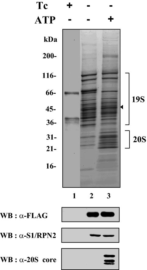
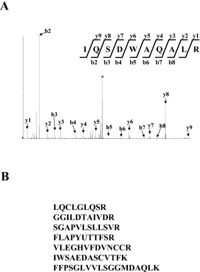
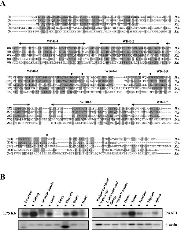
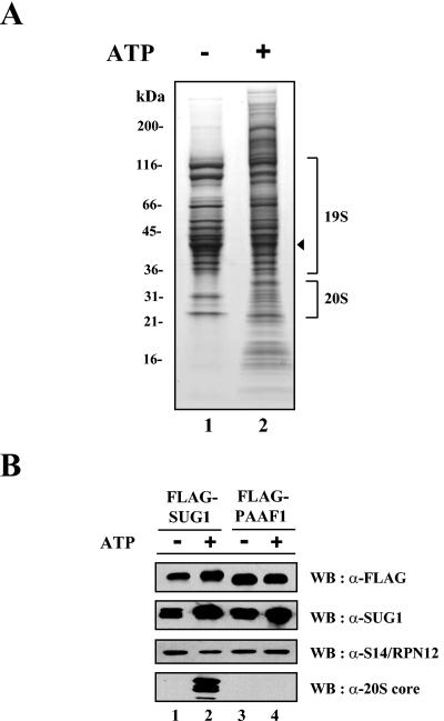
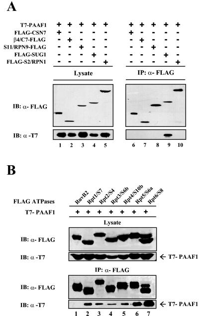
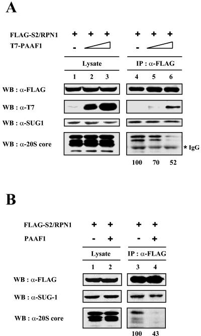
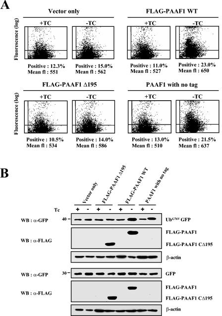
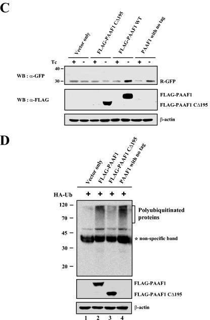
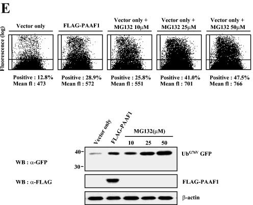
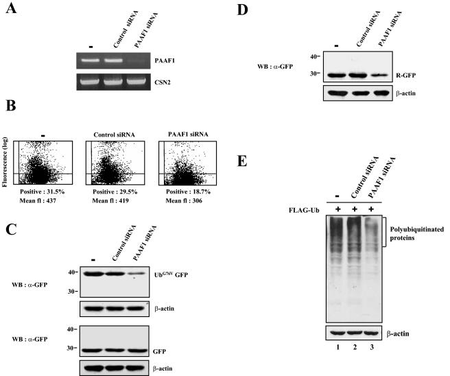
The 26S proteasome, composed of the 20S core and the 19S regulatory complex, plays a central role in ubiquitin-dependent proteolysis by catalyzing degradation of polyubiquitinated proteins. In a search for proteins involved in regulation of the proteasome, we affinity purified the 19S regulatory complex from HeLa cells and identified a novel protein of 43 kDa in size as an associated protein. Immunoprecipitation analyses suggested that this protein specifically interacted with the proteasomal ATPases. Hence the protein was named proteasomal ATPase-associated factor 1 (PAAF1). Immunoaffinity purification of PAAF1 confirmed its interaction with the 19S regulatory complex and further showed that the 19S regulatory complex bound with PAAF1 was not stably associated with the 20S core. Overexpression of PAAF1 in HeLa cells decreased the level of the 20S core associated with the 19S complex in a dose-dependent fashion, suggesting that PAAF1 binding to proteasomal ATPases inhibited the assembly of the 26S proteasome. Proteasomal degradation assays using reporters based on green fluorescent protein revealed that overexpression of PAAF1 inhibited the proteasome activity in vivo. Furthermore, the suppression of PAAF1 expression that is mediated by small inhibitory RNA enhanced the proteasome activity. These results suggest that PAAF1 functions as a negative regulator of the proteasome by controlling the assembly/disassembly of the proteasome.


Figures

FIG. 1.
FIG. 1.
Immunoaffinity purification of FLAG-SUG1. Cells expressing FLAG-SUG1 in a tetracycline-controlled manner were established as described in Materials and Methods. Cells were grown in the presence (+) or absence (−) of tetracycline (Tc). Immunoaffinity purification of FLAG-SUG1 was performed in the presence (+) or absence (−) of 2 mM ATP. Ten microliters each of purified FLAG-SUG1 samples was separated by a SDS-polyacrylamide gel (gradient of 4 to 20% polyacrylamide) and visualized by Sypro Ruby-Coomassie blue double staining. Protein size markers (in kDa) are indicated to the left of the gel. The location of FLAG-SUG1 is indicated by the arrowhead to the right of the gel. The presence of proteasome subunits was analyzed by immunoblotting of the purified samples with the indicated antibodies. WB, Western blotting; α-FLAG, anti-FLAG.
FIG. 2.
FIG. 2.
Identification of PAAF1 in the purified FLAG-SUG1 preparation. (A) MS-MS spectrum of a peptide derived from PAAF1. MS-MS analyses were performed as described in Materials and Methods. The b and y ions detected are marked with arrows on the spectra. The precursor ion is indicated by the asterisk. (B) Sequences determined by MS-MS analyses of tryptic peptides obtained from PAAF1. All peptides were identical to sequences in human PAAF1.
FIG. 3.
FIG. 3.
Sequence alignment and expression pattern of PAAF1. (A) Sequence alignment of human PAAF1 with homologous proteins from other species. Amino acid sequences of PAAF1-related proteins from different species were aligned using ClustalW. Identical residues are shaded gray. WD40 motifs are indicated. Abbreviations and accession numbers of homologous proteins are as follows: Homo sapiens (H.s.), AAH21541; Gallus gallus (G.g.), CAG31874; Xenopus laevis (X.l.), AAH70729; Dictyostelium discoideum (D.d.), AAM43783; Saccharomyces cerevisiae (S.c.), AAT92618. (B) Northern blot analysis of human PAAF1. The blots containing mRNA from the indicated tissues were probed with 32P-labeled PAAF1 cDNA (top blots), stripped, and reprobed with 32P-labeled actin cDNA (bottom blots).
FIG. 4.
FIG. 4.
Immunoaffinity purification of FLAG-PAAF1. (A) SDS-PAGE analysis of purified FLAG-PAAF1. Ten microliters each of FLAG-PAAF1 samples purified in the presence (+) or absence (−) of 2 mM ATP was separated by electrophoresis on an SDS-polyacrylamide gel (gradient of 4 to 20% polyacrylamide) and visualized by Sypro Ruby-Coomassie blue double staining. Protein size markers (in kDa) are indicated to the left of the gel. The location of FLAG-PAAF1 is shown by the arrowhead to the right of the gel. (B) Western blotting (WB) analysis of purified FLAG-PAAF1. FLAG-PAAF1 and FLAG-SUG1 samples purified in the presence (+) or absence (−) of ATP were analyzed by immunoblotting with indicated antibodies. α-FLAG, anti-FLAG.
FIG. 5.
FIG. 5.
PAAF1 interacts with proteasomal ATPases. (A) Immunoprecipitations of proteasome subunits. HeLa cells were transiently transfected with plasmid vectors expressing T7-PAAF1 and a FLAG-tagged proteasome subunit or CSN7A as indicated. Cell lysates were immunoprecipitated (IP) with beads coupled to anti-FLAG antibody (α-FLAG). The immunoprecipitates were subjected to SDS-PAGE and analyzed by Western blotting with indicated antibodies. IB, immunoblotting. (B) Immunoprecipitations of proteasomal ATPases. HeLa cells were transiently transfected with expression constructs for T7-PAAF1 and a FLAG-tagged proteasomal ATPase subunit or RuvB2 as indicated. Lysates were prepared 36 h after transfection and immunoprecipitated (IP) with anti-FLAG (α-FLAG) antibodies. The precipitates were separated by SDS-PAGE and immunoblotted (IB) with the indicated antibodies.
FIG. 6.
FIG. 6.
PAAF1 interferes with the binding of 19S regulatory complex to 20S core. HeLa cells were transiently transfected with FLAG-S2/Rpn1 and T7-tagged (A) or untagged (B) PAAF1. Lysates were prepared 36 h after transfection and immunoprecipitated (IP) with anti-FLAG (α-FLAG) antibodies. The precipitates were separated by SDS-PAGE and immunoblotted with the indicated antibodies. Coprecipitated 20S α-core bands were quantified by Las-3000 luminescent image analyzer (Fuji film). WB, Western blotting; IgG, immunoglobulin G.
FIG. 7.
FIG. 7.
PAAF1 inhibits the proteasome activity. (A) Flow cytometric analysis of UbG76V-GFP. HeLa Tet-off cells (Clontech) were transfected with the expression construct of UbG76V-GFP and with the expression vector for PAAF1 under the control of a tetracycline-inducible promoter as indicated. Cells were incubated in the presence (+) or absence (−) of tetracycline (TC) for 48 h. The GFP fluorescence (fl) was then analyzed by flow cytometry. WT, wild type. (B) Immunoblotting analyses of UbG76V-GFP and GFP. HeLa Tet-off cells were transfected with the expression construct of UbG76V-GFP (top blots) or GFP (bottom blots) and with the expression vector for PAAF1 under the control of a tetracycline-inducible promoter as indicated. After induction of PAAF1 derivatives for 48 h, the expression levels of UbG76V-GFP and GFP were determined by immunoblotting with anti-GFP (α-GFP) antibodies. WB, Western blotting; Tc, tetracycline. (C) Immunoblotting analysis of Ub-R-GFP. HeLa Tet-off cells were transfected with the expression construct of Ub-R-GFP and with the expression vector for PAAF1 under the control of a tetracycline-inducible promoter as indicated. After induction of PAAF1 derivatives for 48 h, the expression levels of Ub-R-GFP were determined by immunoblotting with anti-GFP (α-GFP) antibodies. WB, Western blotting; Tc, tetracycline. (D) Immunoblotting analysis of polyubiquitinated proteins. HeLa cells were transfected with the expression constructs of HA-Ub and FLAG-PAAF1 as indicated. Polyubiqitinated proteins were analyzed by immunoblotting with anti-HA antibodies. The asterisk indicates a nonspecific band detected in whole-cell lysates by anti-HA antibodies. (E) Comparison of the inhibition of proteasome activity by PAAF1 overexpression with that by the proteasome inhibitor MG132. HeLa cells were transfected with the expression construct of UbG76V-GFP and with the expression vector for PAAF1 or the empty vector as indicated. Thirty-six hours after transfection, cells transfected with the empty vector were treated for 10 h with 0, 10, 25, or 50 μM MG132 as indicated. The expression level of UbG76V-GFP was then analyzed by flow cytometry and by immunoblotting with anti-GFP (α-GFP) antibodies. fl, fluorescence; WB, Western blotting.
FIG. 7.
FIG. 7.
PAAF1 inhibits the proteasome activity. (A) Flow cytometric analysis of UbG76V-GFP. HeLa Tet-off cells (Clontech) were transfected with the expression construct of UbG76V-GFP and with the expression vector for PAAF1 under the control of a tetracycline-inducible promoter as indicated. Cells were incubated in the presence (+) or absence (−) of tetracycline (TC) for 48 h. The GFP fluorescence (fl) was then analyzed by flow cytometry. WT, wild type. (B) Immunoblotting analyses of UbG76V-GFP and GFP. HeLa Tet-off cells were transfected with the expression construct of UbG76V-GFP (top blots) or GFP (bottom blots) and with the expression vector for PAAF1 under the control of a tetracycline-inducible promoter as indicated. After induction of PAAF1 derivatives for 48 h, the expression levels of UbG76V-GFP and GFP were determined by immunoblotting with anti-GFP (α-GFP) antibodies. WB, Western blotting; Tc, tetracycline. (C) Immunoblotting analysis of Ub-R-GFP. HeLa Tet-off cells were transfected with the expression construct of Ub-R-GFP and with the expression vector for PAAF1 under the control of a tetracycline-inducible promoter as indicated. After induction of PAAF1 derivatives for 48 h, the expression levels of Ub-R-GFP were determined by immunoblotting with anti-GFP (α-GFP) antibodies. WB, Western blotting; Tc, tetracycline. (D) Immunoblotting analysis of polyubiquitinated proteins. HeLa cells were transfected with the expression constructs of HA-Ub and FLAG-PAAF1 as indicated. Polyubiqitinated proteins were analyzed by immunoblotting with anti-HA antibodies. The asterisk indicates a nonspecific band detected in whole-cell lysates by anti-HA antibodies. (E) Comparison of the inhibition of proteasome activity by PAAF1 overexpression with that by the proteasome inhibitor MG132. HeLa cells were transfected with the expression construct of UbG76V-GFP and with the expression vector for PAAF1 or the empty vector as indicated. Thirty-six hours after transfection, cells transfected with the empty vector were treated for 10 h with 0, 10, 25, or 50 μM MG132 as indicated. The expression level of UbG76V-GFP was then analyzed by flow cytometry and by immunoblotting with anti-GFP (α-GFP) antibodies. fl, fluorescence; WB, Western blotting.
FIG. 8.
FIG. 8.
Proteasome activity is enhanced by siRNA-mediated PAAF1 suppression. (A) siRNA-mediated suppression of PAAF1 mRNA. HeLa cells were transfected with control or PAAF1-specific siRNA as indicated. Total cellular RNA was isolated 48 h after transfection and subjected to RT-PCR analysis using specific primers for PAAF1 and CSN2 mRNA. (B) Flow cytometric analysis of UbG76V-GFP. HeLa cells were transfected with control or PAAF1-specific siRNA as indicated. Twelve hours later, the expression construct for UbG76V-GFP was transfected into siRNA-treated HeLa cells. After incubation for 48 h, the GFP fluorescence (fl) was analyzed by flow cytometry. (C) Immunoblotting analysis of UbG76V-GFP. Transfection experiments were performed as described in the legend to panel B. The expression levels of UbG76V-GFP were determined by immunoblotting with anti-GFP (α-GFP) antibodies. WB, Western blotting. (D) Immunoblotting analysis of Ub-R-GFP. Transfection experiments were performed with the expression construct for Ub-R-GFP as described in the legend to panel B. (E) Immunoblotting analysis of polyubiquitinated proteins. HeLa cells were transfected with control or PAAF1-specific siRNA as indicated. Twelve hours later, the expression construct for FLAG-Ub was transfected into siRNA-treated HeLa cells. After incubation for 48 h, polyubiqitinated proteins were analyzed by immunoblotting with anti-FLAG antibodies.

Similar articles

Cited by

References

    1. Borodovsky, A., B. M. Kessler, R. Casagrande, H. S. Overkleeft, K. D. Wilkinson, and H. L. Ploegh. 2001. A novel active site-directed probe specific for deubiquitylating enzymes reveals proteasome association of USP14. EMBO J. 20:5187-5196. - PMC - PubMed
    1. Boussif, O., F. Lezoualc'h, M. A. Zanta, M. D. Mergny, D. Scherman, B. Demeneix, and J. P. Behr. 1995. A versatile vector for gene and oligonucleotide transfer into cells in culture and in vivo: polyethylenimine. Proc. Natl. Acad. Sci USA 92:7297-7301. - PMC - PubMed
    1. Brown, D. D., Z. Wang, J. D. Furlow, A. Kanamori, R. A. Schwartzman, B. F. Remo, and A. Pinder. 1996. The thyroid hormone-induced tail resorption program during Xenopus laevis metamorphosis. Proc. Natl. Acad. Sci. USA 93:1924-1929. - PMC - PubMed
    1. Chomczynski, P., and N. Sacchi. 1987. Single-step method of RNA isolation by acid guanidinium thiocyanate-phenol-chloroform extraction. Anal. Biochem. 162:156-159. - PubMed
    1. Dantuma, N. P., K. Lindsten, R. Glas, M. Jellne, and M. G. Masucci. 2000. Short-lived green fluorescent proteins for quantifying ubiquitin/proteasome-dependent proteolysis in living cells. Nat. Biotechnol. 18:538-543. - PubMed

Publication types

MeSH terms

Substances

LinkOut - more resources