. 2005 Aug;19(8):2035-46.
doi: 10.1210/me.2004-0531. Epub 2005 Apr 14.

The modulator of nongenomic actions of the estrogen receptor (MNAR) regulates transcription-independent androgen receptor-mediated signaling: evidence that MNAR participates in G protein-regulated meiosis in Xenopus laevis oocytes

Affiliations

The modulator of nongenomic actions of the estrogen receptor (MNAR) regulates transcription-independent androgen receptor-mediated signaling: evidence that MNAR participates in G protein-regulated meiosis in Xenopus laevis oocytes

Derek Haas et al. Mol Endocrinol. 2005 Aug.

Abstract

Classical steroid receptors mediate many transcription-independent (nongenomic) steroid responses in vitro, including activation of Src and G proteins. Estrogen-triggered activation of Src can be regulated by the modulator of nongenomic actions of the estrogen receptor (MNAR), which binds to estrogen receptors and Src to create a signaling complex. In contrast, the mechanisms regulating steroid-induced G protein activation are not known, nor are the physiologic responses mediated by MNAR. These studies demonstrate that MNAR regulates the biologically relevant process of meiosis in Xenopus laevis oocytes. MNAR was located throughout oocytes, and reduction of its expression by RNA interference markedly enhanced testosterone-triggered maturation and activation of MAPK. Additionally, Xenopus MNAR augmented androgen receptor (AR)-mediated transcription in CV1 cells through activation of Src. MNAR and AR coimmunoprecipitated as a complex involving the LXXLL-rich segment of MNAR and the ligand binding domain of AR. MNAR and Gbeta also precipitated together, with the same region of MNAR being important for this interaction. Finally, reduction of MNAR expression decreased Gbetagamma-mediated signaling in oocytes. MNAR therefore appears to participate in maintaining meiotic arrest, perhaps by directly enhancing Gbetagamma-mediated inhibition of meiosis. Androgen binding to AR might then release this inhibition, allowing maturation to occur. Thus, MNAR may augment multiple nongenomic signals, depending upon the context and cell type in which it is expressed.


Figures

Fig. 1
Fig. 1
Protein Sequence of XeMNAR The 1012 amino acid coding sequence of XeMNAR is depicted. The seven LXXLL motifs (potential steroid receptor binding sequences) are boxed, and the proline-rich motifs (potential SH3 or WW binding domains) underlined. The truncated MNAR523stop protein contains a stop codon just after the lysine residue at position 522 (circled).
Fig. 2
Fig. 2
MNAR Is Expressed throughout Xenopus laevis Oocytes Immunohistochemistry was performed on oocyte sections as described in Materials and Methods. For either the top set or bottom set of samples, oocytes were incubated for an equal amount of time with equal concentrations of protein A-purified rabbit antibodies from the serum of rabbits injected with a peptide containing amino acids 991–1007 (immune) or from the corresponding preimmune serum (preimmune). Normal oocytes (uninjected, top) or oocytes injected with double-stranded cRNA corresponding to XeMNAR (dsXeMNAR, bottom) were used. Brown staining depicts expression. More than five sections from five different oocytes were examined for each condition with identical results.
Fig. 3
Fig. 3
XeMNAR Enhances Androgen-Triggered XeAR-Mediated Transcription A and B, XeAR-mediated transcription was measured in CV1 cells treated for approximately 48 h with either testosterone (A) or DHT (B). Cells were cotransfected with MMTV-luciferase, CMV-galactosidase, XeAR, and either empty pcDNA3.1 vector (open squares) or pcDNA3.1 containing a cDNA encoding Myc-tagged XeMNAR (closed circles). Steroid concentrations are depicted on the x-axis (log scale) and luciferase activity normalized to CMV promoter-driven β-galactosidase production on the y-axis. C, XeMNAR’s enhancement of AR-mediated transcription is regulated via Src. CV1 cells were transfected as above, but were treated with either ethanol vehicle or the Src inhibitor PP2 (a gift from William Rainey, University of Texas Southwestern) at a concentration of 4 μM during the 48-h incubation with 10 nM DHT. Each point in each experiment represents the average ± SD (n = 3), and each experiment was performed at least two times with nearly identical results.
Fig. 4
Fig. 4
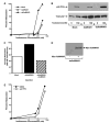
Endogenous MNAR Blocks Meiotic Progression in Xenopus laevis Oocytes A, Reduction of endogenous MNAR expression in oocytes by RNA interference enhances testosterone-triggered maturation. Xenopus oocytes were injected with either buffer (mock, closed circles), cRNA encoding XeMNAR (open squares), or double-stranded cRNA corresponding the XeMNAR (closed triangles). After 36 h, oocytes were treated overnight with testosterone at the indicated concentrations (x-axis) and maturation determined by detection of germinal vesicle breakdown (percent maturation, y-axis, n = 20 per point). This experiment was performed more than five times with nearly identical results. B, Oocytes were injected as in panel A. After 48 h, oocytes were treated with either ethanol vehicle or 150 nM testosterone for 4 h, and phosphorylated p42 ERK detected by Western blot (upper panel). The blot was then stripped and reprobed for total p42 (lower panel). Phosphorylation of p42 ERK is significantly higher in oocytes injected with dsXeMNAR. This experiment was repeated three times with nearly identical results. C, Oocytes were injected with either buffer (Mock) or double-stranded cRNA corresponding to XeMNAR (dsXeMNAR). The latter oocytes were then injected with single-stranded cRNA encoding Myc-XeMNAR 24 h later. Oocyte maturation was examined as above 24 h later by treating oocytes overnight with 300 nM testosterone. The Myc-tagged XeMNAR was indeed overexpressed in the double-injected cells, as indicated by Western blot using a mouse monoclonal anti-Myc antibody (D). E, Inhibition of Src has minimal effect on testosterone-induced oocyte maturation. Uninjected oocytes were preincubated with either dimethylsulfoxide (DMSO) vehicle (closed circle) or 4 μM PP2 (open square) for 2 h followed by the addition of testosterone at the indicated concentrations. Maturation was determined 16 h later and is indicated on the y-axis.
Fig. 5
Fig. 5
Endogenous XeMNAR Coprecipitates with XeAR and XeGβ in Oocyte Extracts Lysates from mock and testosterone-treated oocytes were prepared and immunoprecipitations of XeAR (left) and XeGβ (right) were performed as described in Materials and Methods. Samples were precleared with preimmune antibody followed by precipitation with the specific immune antibodies (immunoprecipitation, IP). All precipitates were then blotted with anti-XeMNAR serum directed against residues 430–447. This experiment was repeated twice with similar results. In addition, the same MNAR band was identified when precipitates were blotted with the anti-XeMNAR serum directed against residues 991–1007.
Fig. 6
Fig. 6
XeMNAR Coprecipitates with XeAR in COS Cells COS cells were transfected with a total of 1 μg DNA that consisted of either pcDNA3.1 alone or pcDNA3.1 containing cDNAs encoding Myc-tagged XeMNAR (0.5 μg) or Myc-tagged XeAR (0.5 μg). Transfection was followed 48 h later by coimmunoprecipitation as described in Materials and Methods. Lysates were precleared with preimmune antibodies followed by immunoprecipitation using rabbit polyclonal antibodies directed against either the XeAR or XeMNAR. Blots were then probed with a monoclonal mouse anti-Myc antibody. Precipitations using the immune sera are shown in the top panels (IP, immunoprecipitation), whereas precipitations using the preimmune sera are shown in the lower panels. Total lysates were probed as positive controls for the Western blots, and antibodies used for the precipitations are indicated. A, XeMNAR coprecipitates with XeAR both in the absence (left) and presence (right) of testosterone. B, XeAR coprecipitates with XeMNAR with and without testosterone. C, Cells were transfected with a cDNA encoding a Myc-tagged truncated XeMNAR (MNAR523stop) instead of full-length XeMNAR. MNAR523stop still coprecipitated with XeAR as indicated. D, Cells were transfected with a cDNA encoding a Myc-tagged truncated XeAR (AR527stop) instead of full-length XeAR. AR527stop did not coprecipitate with XeMNAR. All experiments were repeated at least three times with identical results.
Fig. 7
Fig. 7
XeMNAR Coprecipitates with Gβin COS Cells A, COS cells were transfected with a total of 1 μg DNA that consisted of either pcDNA3.1 alone or pcDNA3.1 containing cDNAs encoding either Myc-tagged XeMNAR or Myc-tagged XeGβ. Transfection was followed 48 h later by coimmunoprecipitation as described in Materials and Methods. Lysates were precleared with nonspecific preimmune antibodies followed by immunoprecipitation using rabbit polyclonal antibodies directed against XeMNAR (IP, immunoprecipitation). Blots were then probed with a monoclonal mouse anti-Myc antibody. Precipitations using the immune sera are shown in the top panels, whereas precipitations using the nonspecific preimmune sera are shown in the lower panels. Total lysates were probed as positive controls and precipitates are indicated. B, COS cells were transfected with the indicated plasmids and complexes were precipitated with rabbit anti-Gβ serum or nonspecific preimmune sera. Blots were probed with the anti-Myc monoclonal antibody. XeMNAR was precipitated with endogenous Gβ. The ability of the anti-Gβ antibody to precipitate Gβ was confirmed by overexpression of Myc-XeGβ in oocytes followed by precipitation with the anti-Gβ antibody (far right lane). C, COS cells were transfected as indicated and precipitations performed using the polyclonal antibodies directed against residues 430–447 of MNAR. Myc-tagged MNAR523stop and XeGβ were detected by Western blot. All experiments were repeated at least three times with identical results.
Fig. 8
Fig. 8
MNAR Mediates Gβγ Signaling A, Reduction of endogenous XeMNAR expression by RNA interference attenuates Gβγ-mediated signaling. Oocytes injected with cRNA encoding the M2R alone (left) or with M2R cRNA plus double-stranded cRNA corresponding to XeMNAR (dsMNAR) were preincubated with 45Ca followed by treatment for the indicated times with 30 μM carbachol. Calcium efflux, which reflects Gβγ signaling via phospholipase Cβ, was measured and presented as fold-induction over baseline on the y-axis. Each bar represents the average of three pools of 10 oocytes ± SD. This experiment was repeated twice with similar results. B, Schematic diagram depicting a possible role of MNAR in maintaining meiotic arrest. The LXXLL, proline-rich (P-Rich), and acidic domains of MNAR are indicated, as are the A/B, DNA binding (DNABD), and ligand binding (LBD) domains of AR. XeMNAR forms a complex with XeAR and XeGβγ (and possibly Gαs) that involves the amino-terminal LXXLL-containing half of XeMNAR to increase G protein activity and maintain meiotic arrest. Androgen binding leads to a change in the conformation of the complex (perhaps without altering the binding of XeAR to XeMNAR) to attenuate G protein-mediated signaling and permit meiosis to resume.

Similar articles

Cited by

References

    1. Cato AC, Nestl A, Mink S. Rapid actions of steroid receptors in cellular signaling pathways. Sci STKE. 2002;2002:RE9. - PubMed
    1. Edwards DP. Regulation of signal transduction pathways by estrogen and progesterone. Annu Rev Physiol. 2005;67:335–376. - PubMed
    1. Boonyaratanakornkit V, Scott MP, Ribon V, Sherman L, Anderson SM, Maller JL, Miller WT, Edwards DP. Progesterone receptor contains a proline-rich motif that directly interacts with SH3 domains and activates c-Src family tyrosine kinases. Mol Cell. 2001;8:269–280. - PubMed
    1. Pietras RJ, Szego CM. Specific binding sites for oestrogen at the outer surfaces of isolated endometrial cells. Nature. 1977;265:69–72. - PubMed
    1. Shaul PW. Rapid activation of endothelial nitric oxide synthase by estrogen. Steroids. 1999;64:28–34. - PubMed

Publication types

MeSH terms