. 2005 Aug 1;106(3):996-1002.
doi: 10.1182/blood-2005-02-0707. Epub 2005 Apr 14.

Crystal structure of the Jak3 kinase domain in complex with a staurosporine analog

Affiliations

Crystal structure of the Jak3 kinase domain in complex with a staurosporine analog

Titus J Boggon et al. Blood. .

Abstract

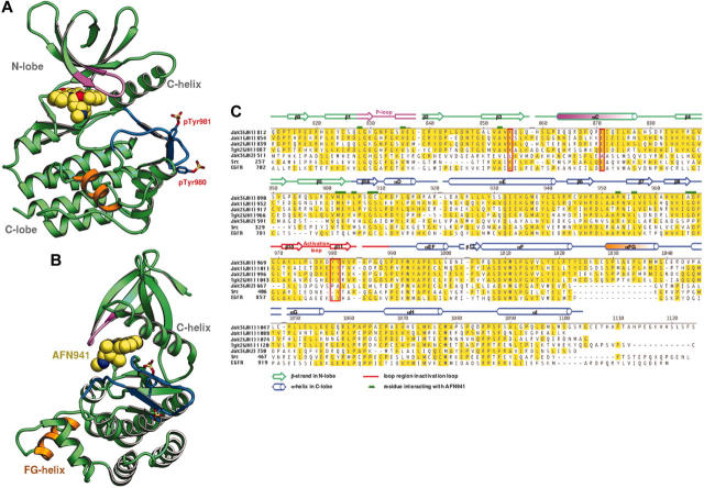
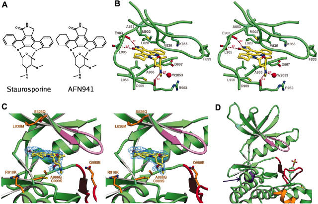
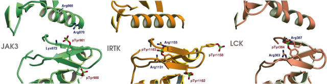
Jak (Janus kinase) family nonreceptor tyrosine kinases are central mediators of cytokine signaling. The Jak kinases exhibit distinct cytokine receptor association profiles and so transduce different signals. Jak3 expression is limited to the immune system, where it plays a key role in signal transduction from cytokine receptors containing the common gamma-chain, gammac. Patients unable to signal via gammac present with severe combined immunodeficiency (SCID). The finding that Jak3 mutations result in SCID has made it a target for development of lymphocyte-specific immunosuppressants. Here, we present the crystal structure of the Jak3 kinase domain in complex with staurosporine analog AFN941. The kinase domain is in the active conformation, with both activation loop tyrosine residues phosphorylated. The phosphate group on pTyr981 in the activation loop is in part coordinated by an arginine residue in the regulatory C-helix, suggesting a direct mechanism by which the active position of the C-helix is induced by phosphorylation of the activation loop. Such a direct coupling has not been previously observed in tyrosine kinases and may be unique to Jak kinases. The crystal structure provides a detailed view of the Jak3 active site and will facilitate computational and structure-directed approaches to development of Jak3-specific inhibitors.


Figures

Figure 1.
Figure 1.
Ribbon diagram of the Jak3 kinase domain and sequence alignment of Jak family members. (A) Overview of the crystal structure of the catalytic domain of Jak3 in complex with tetrahydro-staurosporine AFN941. β-Strands are shown as arrows and α-helices as coils. The glycine-rich loop is colored purple and the activation-loop is blue. The novel αFG helix is colored orange. The N-lobe, C-lobe, and C-helix are indicated. Phosphorylated tyrosines pTyr980 and pTyr981 are shown in stick form. The staurosporine analog is shown as space-filling spheres with carbon atoms colored yellow. (B) Ribbon diagram as in panel A but with the view rotated by 90°. (C) Sequence alignment for the kinase (JH1) domains of Jak3, Jak1, Jak2, and Tyk2; the JH2 domain of Jak3; and the kinase domains of c-Src and epidermal growth factor receptor (EGFR). The numbering and secondary structure assignments for Jak3 crystal structure are indicated. β-Strands are indicated as arrows, α-helices as cylinders, and loops as lines. The activation loop is indicated in red, and the novel helix αFG in orange. The N-lobe is denoted by green secondary structure assignments and the C-lobe with blue. Residues that interact with the staurosporine analog AFN941 are indicated by green boxes. The catalytically important residues Lys855, Glu871 are boxed in red as are the phosphorylated tyrosine residues pTyr980 and pTyr981. GenBank accession codes for Jak1, Jak2, Jak3, Tyk2, c-Src, and EGFR are NP_002218, NP_004963, NP_000206, AAS37680, AAH11566, and NP_005219, respectively.
Figure 2.
Figure 2.
Binding of AFN941 to Jak3 active site. (A) Structure of AFN941 juxtaposed with that of staurosporine. (B) Stereoview of the catalytic cleft of Jak3 bound to AFN941. The protein backbone trace is shown in the region of the active site with residues Leu828, Phe833, Val836, Ala853, Lys855, Met902, Glu903, Cys909, Arg953, Leu956, Ala966, and Asp967 depicted in stick representation. Staurosporine analog AFN941 is shown in stick representation with carbon atoms colored yellow. Carbonyl oxygens of residues Arg953 and Glu903 are shown in red, and water 2053 is depicted as a red sphere. Residue numbers are indicated. Hydrogen bonds to backbone atoms and water are indicated and their distances are noted. (C) Stereoview of the catalytic cleft of Jak3 bound to the staurosporine analog AFN941. The experimental Fobs - Fcalc electron density omit map contoured at 2.5σ in blue is shown for the ligand AFN941. The glycine-rich loop is shown in purple and the activation loop in red. Residues proximal to the active site and divergent between Jak3 and Jak2 are shown in orange stick format and are labeled by Jak3 residue type, Jak3 residue number, and Jak2 residue type. These residues are Ser828Gln, Leu838Met, Cys909Ser, Arg916Lys, Ala966Gly, and Gln988Glu. (D) Location of point mutation Leu910Ser noted in human SCID patients. Leu910 is colored purple and indicated by the arrow.
Figure 3.
Figure 3.
Comparison of activation loop conformation for Jak3, Lck, and IRTK. The 3 panels show ribbon representation diagrams for the activation loops of Jak3, IRTK,31 and Lck. The Jak3 activation loop is phosphorylated on 2 adjacent tyrosines, Tyr980 and Tyr981. pTyr981 is in a similar location to phosphorylated tyrosines in the previously determined activated crystal structures of Lck (PDB entry 3LCK) and insulin receptor tyrosine kinase31 (PDB entry 1IR3). Lck is phosphorylated on a single tyrosine, in the equivalent position to Tyr981; however, insulin receptor is phosphorylated on 3 tyrosines on the activation loop, 2 of which are the equivalent residues to Tyr980 and Tyr981 in Jak3. pTyr980 in Jak3 is in a similar conformation to that of pTyr1162 in IRTK. In Jak3, Arg870 of the C-helix coordinated pTyr981. Arg866 also extends toward pTyr981 but is poorly ordered; therefore, its side chain is not illustrated. Figures were prepared using the program SETOR.

References

    1. Rane SG, Reddy EP. Janus kinases: components of multiple signaling pathways. Oncogene. 2000;19: 5662-5679. - PubMed
    1. Imada K, Leonard WJ. The Jak-STAT pathway. Mol Immunol. 2000;37: 1-11. - PubMed
    1. Saharinen P, Takaluoma K, Silvennoinen O. Regulation of the Jak2 tyrosine kinase by its pseudokinase domain. Mol Cell Biol. 2000;20: 3387-3395. - PMC - PubMed
    1. O'Shea JJ, Husa M, Li D, et al. Jak3 and the pathogenesis of severe combined immunodeficiency. Mol Immunol. 2004;41: 727-737. - PubMed
    1. Buckley RH, Schiff RI, Schiff SE, et al. Human severe combined immunodeficiency: genetic, phenotypic, and functional diversity in one hundred eight infants. J Pediatr. 1997;130: 378-387. - PubMed

Publication types