Comparative Study
. 2005 Apr 15;6(1):36.
doi: 10.1186/1465-9921-6-36.

Inhibitors of mitogen-activated protein kinases differentially regulate costimulated T cell cytokine production and mouse airway eosinophilia

Affiliations
Comparative Study

Inhibitors of mitogen-activated protein kinases differentially regulate costimulated T cell cytokine production and mouse airway eosinophilia

Ligia Chialda et al. Respir Res. .

Abstract

Background: T cells play a dominant role in the pathogenesis of asthma. Costimulation of T cells is necessary to fully activate them. An inducible costimulator (ICOS) of T cells is predominantly expressed on Th2 cells. Therefore, interference of signaling pathways precipitated by ICOS may present new therapeutic options for Th2 dominated diseases such as asthma. However, these signaling pathways are poorly characterized in vitro and in vivo.

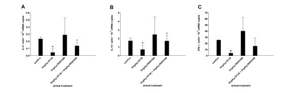
Methods: Human primary CD4+ T cells from blood were activated by beads with defined combinations of surface receptor stimulating antibodies and costimulatory receptor ligands. Real-time RT-PCR was used for measuring the production of cytokines from activated T cells. Activation of mitogen activated protein kinase (MAPK) signaling pathways leading to cytokine synthesis were investigated by western blot analysis and by specific inhibitors. The effect of inhibitors in vivo was tested in a murine asthma model of late phase eosinophilia. Lung inflammation was assessed by differential cell count of the bronchoalveolar lavage, determination of serum IgE and lung histology.

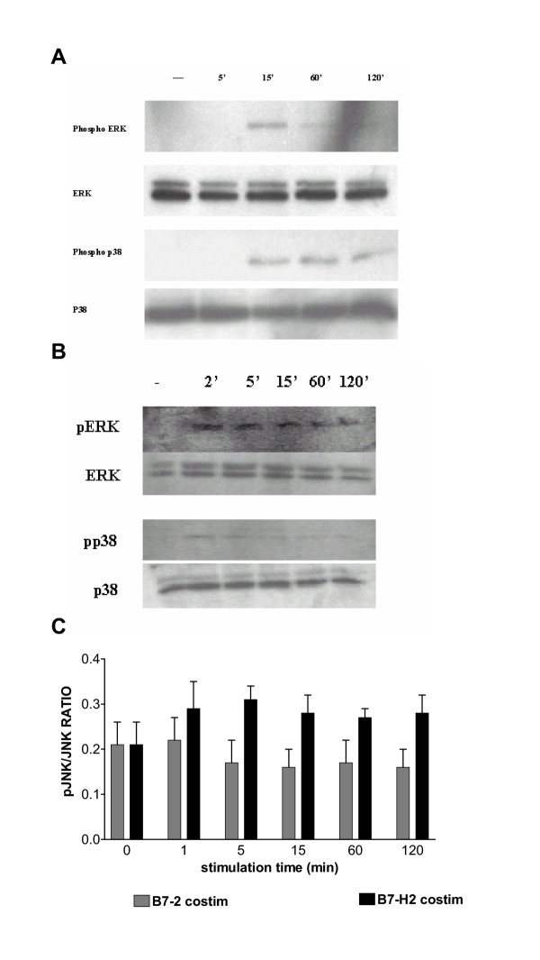
Results: We showed in vitro that ICOS and CD28 are stimulatory members of an expanding family of co-receptors, whereas PD1 ligands failed to co-stimulate T cells. ICOS and CD28 activated different MAPK signaling cascades necessary for cytokine activation. By means of specific inhibitors we showed that p38 and ERK act downstream of CD28 and that ERK and JNK act downstream of ICOS leading to the induction of various T cell derived cytokines. Using a murine asthma model of late phase eosinophilia, we demonstrated that the ERK inhibitor U0126 and the JNK inhibitor SP600125 inhibited lung inflammation in vivo. This inhibition correlated with the inhibition of Th2 cytokines in the BAL fluid. Despite acting on different signaling cascades, we could not detect synergistic action of any combination of MAPK inhibitors. In contrast, we found that the p38 inhibitor SB203580 antagonizes the action of the ERK inhibitor U0126 in vitro and in vivo.

Conclusion: These results demonstrate that the MAPKs ERK and JNK may be suitable targets for anti-inflammatory therapy of asthma, whereas inhibition of p38 seems to be an unlikely target.


Figures

Figure 1
Figure 1
Induction of cytokines in CD4+ T cells by different costimulatory receptors. Human CD4+ T cells were stimulated with various stimuli. (A) Cytokine mRNA level were determined after eight hours stimulation using real-time RT-PCR. mRNA level were normalized to β-actin and CD3 stimulated cells were set to 1. Each column represents mean ± s.e.mean of three different donors. (B) Cytokine protein level was determined in the supernatant by ELISA after twenty-four hours of stimulation. Each column represents mean ± s.e.mean of three different volunteers. *P < 0.05 (versus CD3 stimulated samples).
Figure 2
Figure 2
Time course of cytokine induction by costimulatory receptors. Human CD4+ T cells were stimulated by α-CD3/B7-2 (A) and α-CD3/B7-H2 (B) respectively and harvested after different time points. Cytokine mRNA level were determined using real-time RT-PCR. Levels were normalized to β-actin and unstimulated cells were set to 1. Each point and bar represents mean ± s.e.mean of duplicates measurements of one donor. Similar results were obtained from three independent donors.
Figure 3
Figure 3
Activation of MAPKs in human primary CD4+ T-cells by costimulatory receptors. Human CD4+ T cells were seeded at a concentration of 1 × 10⁶/ml and stimulated with Dynal beads coated with α-CD3/B7-2 Fc (A) or α-CD3/B7-H2 Fc (B) for 0, 2, 5, 15, 60 or 120 min as indicated. Cells were harvested and cell extracts prepared. Cell extracts were analyzed by Western blot using either Anti-active pAb (upper row) or with corresponding antibodies that recognize both active and inactive forms of each subfamily of MAPK. Results are representative of three experiments with similar results. (C) Activated JNK was detected by ELISA. CD4+ T cells were cultured in precoated 96-well plates at a concentration of 1 × 10⁶ /ml. Cells were stimulated with α-CD3/B7-2 Fc and α-CD3/B7-H2 Fc for different times (1, 5, 15, 60 and 120 min). Following stimulation the cells were fixed. The phosphorylated form of JNK as well as the amount of total JNK was detected using an ELISA method. Results shown are representative for three independent experiments.
Figure 4
Figure 4
Effects of MAPK inhibitors in vivo. Actively intraperitoneally sensitized mice were challenged by intranasally administration of ovalbumin. SB203580 (Figure A), SP600125 (Figure B) and U0126 (Figure C) were given intraperitoneally two hours prior to ovalbumin challenge. After 4 days of challenge bronchoalveolar lavage was performed. Total cell number in BAL and differential cell count on cytospin preparations was assessed. OVA challenged vehicle-treated animals were taken as a positive control. Each column represents mean ± s.e.mean. n = 5 mice per group. *p < 0.05 (drug treated versus OVA challenged vehicle (positive) control), **p < 0.005 (drug treated versus OVA challenged vehicle (positive) control). ++ p < 0.005 (OVA challenged (positive) versus saline challenged (negative) control).
Figure 5
Figure 5
Effects of MAPK inhibitors on BAL cytokine levels. Cells were recovered from bronchoalveolar lavage by centrifugation and RNA was prepared from the cell pellet. Cytokine mRNA level were determined using real-time RT-PCR and were normalized to β-actin. Each column represents mean ± s.e.mean. n = 5 mice per group. n.d. – not detectable. *p < 0.05 (versus OVA challenged positive control), +p < 0.05 (negative versus positive control)
Figure 6
Figure 6
Effects of MAPK inhibitors on OVA-specific IgE antibody levels in the serum. Mouse serum was collected 24 h after the last OVA challenge. Samples were obtained from saline- (negative) or OVA-challenged mice in the absence (positive) or presence of MAPK inhibitors: SB203580 (20 mg/kg), U0126 (20 mg/kg), SP600125 (20 mg/kg) or of the combination of U0126 (5 mg/kg) plus SB203580 (5 mg/kg). Serum titers for OVA-specific IgE antibodies were measured by ELISA as described in methods. Each column represents mean ± s.e.mean. n = 5 mice per group. *p < 0.05 (versus OVA challenged positive control), +p < 0.05 (negative versus positive control)
Figure 7
Figure 7
Histology of lung inflammation. Representative sections of the lungs are shown. Sections are periodic acid-Schiff stained and counterstained with haematoxylin for analysis of mucin-containing cells. Tissue was examined by light microscopy (original magnification 20×), zoomed inset delineates PAS+ epithelial cells (100×). Lung sections were obtained from saline- (A) or OVA- (B, C, D, E, F) challenged mice in the absence (A, B) or presence of MAPK inhibitors: SB 203580 (20 mg/kg) (C), U 0126 (20 mg/kg) (D), SP 600125 (20 mg/kg) (E) or of the combination of U 0126 (5 mg/kg) plus SB 203580 (5 mg/kg) (F). Peribronchial inflammation and the abundance of PAS-positive mucus-containing cells were determined by applying the scores as described in methods (G). The scores described in methods were applied to ten bronchi for each animal and mean and standard deviation were determined for all the animals in a group (n = 5). *P < 0.05 and **P < 0.01 compared with positive control group.
Figure 8
Figure 8
Effects of combined administration of MAPK inhibitors in vitro. Human CD4+ T cells were preincubated either with increasing doses of U0126 alone (solid line) or in combination with SP 600125 (dashed line) of with SB 203580 (dotted line). Doses of the MAPK inhibitors are specitied in the text. After that, cells were stimulated with α-CD3/B7-2 Fc (A, B) or α-CD3/B7-H2 Fc (C, D) for 8 hours. IFNγ (A, C) and IL-13 (B, D) cytokine mRNA was measured by real-time RT-PCR. Dose-response curves were determined and IC50 values were calculated. Data are representative of three different donors.
Figure 9
Figure 9
Effects of combined administration of MAPK inhibitors in vivo. Late phase eosinophilia was induced in mice pretreated either with 5 mg/kg U0126 and 5 mg/kg SB 203580 or with the combination thereof. BAL was performed and total cell number and differential leukocyte count was determined. OVA challenged vehicle-treated animals were taken as positive control. Each column represents mean ± s.e.mean. n = 5 mice per group. *p < 0.05 (versus OVA challenged vehicle control), +p < 0.05 (versus U0126 alone).
Figure 10
Figure 10
Effects of combined MAPK inhibitors on BAL cytokine levels. Cells were recovered from bronchoalveolar lavage by centrifugation and RNA was prepared from the cell pellet. Cytokine mRNA level were determined using real-time RT-PCR and were normalized to β-actin. Each column represents mean ± s.e.mean. n = 5 mice per group. *p < 0.05 (versus OVA challenged vehicle control), +p < 0.05 (versus U0126 alone).

References

    1. Busse WW, Lemanske RFJ. Asthma. N Engl J Med. 2001;344:350–362. doi: 10.1056/NEJM200102013440507. - DOI - PubMed
    1. Chung KF, Barnes PJ. Cytokines in asthma. Thorax. 1999;54:825–857. - PMC - PubMed
    1. Yssel H, Groux H. Characterization of T cell subpopulations involved in the pathogenesis of asthma and allergic diseases. Int Arch Allergy Immunol. 2000;121:10–18. doi: 10.1159/000024292. - DOI - PubMed
    1. Ray A, Cohn L. Altering the Th1/Th2 balance as a therapeutic strategy in asthmatic diseases. Curr Opin Investig Drugs. 2000;1:442–448. - PubMed
    1. Pahl A, Szelenyi I. Asthma therapy in the new millennium. Inflamm Res. 2002;51:273–282. - PubMed

Publication types