. 2005 Apr 26;102(17):6183-8.
doi: 10.1073/pnas.0502070102. Epub 2005 Apr 15.

Corticotropin-releasing hormone activates ERK1/2 MAPK in specific brain areas

Affiliations

Corticotropin-releasing hormone activates ERK1/2 MAPK in specific brain areas

Damián Refojo et al. Proc Natl Acad Sci U S A. .

Abstract

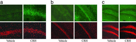
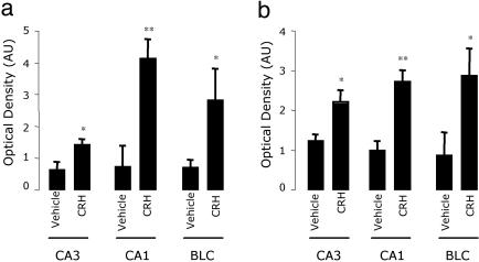
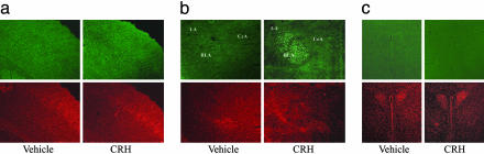
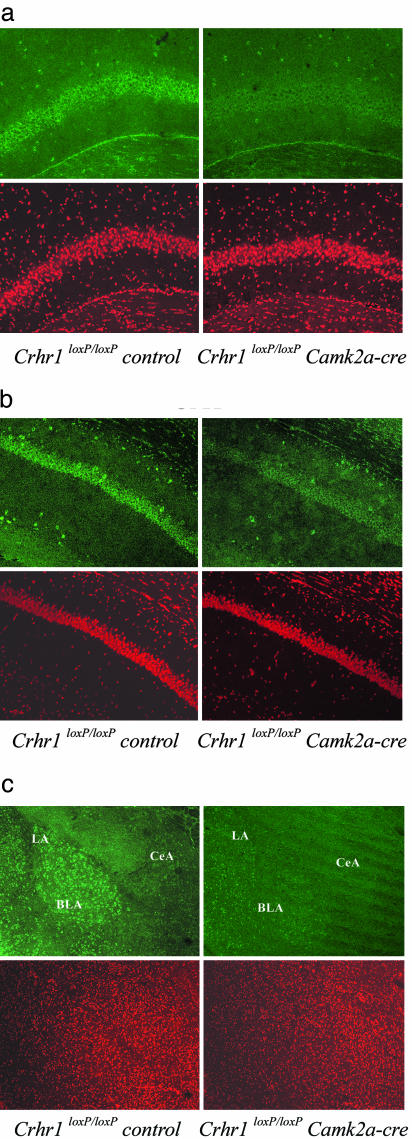
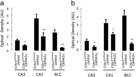
Corticotropin-releasing hormone (CRH) coordinates hormonal and behavioral responses to stress. The mitogen-activated protein kinase extracellular signal-related kinase 1/2 (ERK1/2) mediates several functions in different forebrain structures and recently has been implicated in CRH signaling in cultured cells. To study in vivo CRH-mediated activation of central ERK1/2, we investigated the expression pattern of the phosphorylated ERK1/2(p-ERK1/2) in the mouse brain after intracerebroventricular CRH injections. As shown by immunohistochemistry and confocal microscopy analysis, CRH administration increased p-ERK1/2 levels specifically in the CA3 and CA1 hippocampal subfields and basolateral complex of the amygdala, both structures related to external environmental information processing and behavioral aspects of stress. Other regions such as hypothalamic nuclei and the central nucleus of the amygdala, also related to central CRH system but involved in the processing of the ascending visceral information and neuroendocrine-autonomic response to stress, did not show CRH-mediated ERK1/2 activation. To dissect the involvement of CRH receptor 1 (CRHR1) and CRHR2, we used conditional knockout mice in which Crhr1 is inactivated in the anterior forebrain and limbic structures. The conditional genetic ablation of Crhr1 inhibited the p-ERK1/2 increase, underlining the involvement of CRHR1 in the CRH-mediated activation. These findings underscore the fact that CRH activates p-ERK1/2 through CRHR1 only in selected brain regions, pointing to a specific role of this pathway in mediating behavioral adaptation to stress.


Figures

Fig. 1.
Fig. 1.
Differential activation of p-ERK1/2-ir in hippocampal neurons after vehicle or i.c.v. CRH injections. Representative images (from five mice) of p-ERK1/2 immunohistochemistry (Upper) and control PI staining (Lower) obtained as described in Methods. Pyramidal cells of the CA3 (a) and CA1 (b) subfields strongly express p-ERK1/2 30 min after i.c.v. CRH injections. In contrast, the dentate gyrus (c) does not express basal or CRH-induced p-ERK1/2 levels. (Magnification: a, ×400; b and c, ×200.)
Fig. 3.
Fig. 3.
Quantitative analysis of p-ERK1/2-ir. Levels of p-ERK1/2 in CA3, CA1, and the BLC (BLC = LA and BLA) 10 min (a) and 30 min (b) after i.c.v. vehicle or CRH administration. Optical density data processed with the fluoview acquisition/analyzer program are shown as optical density arbitrary units (AU) after subtraction of each image with its corresponding background signal. Data are presented as mean ± SE, n = 5 animals; *, P < 0.05; **, P < 0.01 vs. the corresponding control vehicle, using ANOVA with Scheffé's test.
Fig. 2.
Fig. 2.
CRH induces p-ERK1/2 levels in the amygdala but not in the cortex or hypothalamus. Representative images (from five mice) of p-ERK1/2 immunohistochemistry (Upper) and control PI staining (Lower) obtained 30 min after i.c.v. CRH injections as described in Methods. (a) Cortex presents moderate levels of basal p-ERK1/2 activation in layers III and IV and CRH does not induce changes. (b) CRH strongly activates ERK1/2 in the LA and BLA. The central amygdaloid nucleus does not present basal or CRH-induced p-ERK1/2 levels. (c) The PVN of the hypothalamus remain absent of any p-ERK1/2 signal in both basal and stimulated conditions. (Magnification: ×200.)
Fig. 4.
Fig. 4.
Crhr1loxP/loxPCamk2a-cre mice show a decreased ERK1/2 activation response in CA3, CA1, and the BLC after central CRH administration. Representative images (from five mice) of p-ERK1/2 immunohistochemistry (Upper) and control PI staining (Lower) obtained 30 min after i.c.v. CRH injections as described in Methods. ERK1/2 activation is greatly reduced in CA3 (a), CA1 (b), and the BLA (c) of CRH-treated Crhr1loxP/loxPCamk2a-cre compared with CRH-treated Crhr1loxP/loxP control mice. (Magnification: ×200.)
Fig. 5.
Fig. 5.
Quantitative analysis of p-ERK1/2-ir in Crhr1loxP/loxPcontrol and Crhr1loxP/loxPCamk2a-cre mice after central CRH administration. Levels of p-ERK1/2 in CA3, CA1, and the BLC (BLC = LA and BLA) of the amygdala 10 min (a) and 30 min (b) after i.c.v. CRH administration. Optical density data processed with the fluoview acquisition/analyzer program are shown as optical density arbitrary units (AU) after subtraction of each image with its corresponding background signal. Data are presented as mean ± SE, n = 5 animals; *, P < 0.05; **, P < 0.01 vs. the corresponding Crhr1loxP/loxPcontrol mice, using ANOVA with Scheffé's test.

References

    1. Sawchenko, P. E., Brown, E. R., Chan, R. K., Ericsson, A., Li, H. Y., Roland, B. L. & Kovacs, K. J. (1996) Prog. Brain Res. 107, 201-222. - PubMed
    1. Holsboer, F. (1999) J. Psychiatr. Res. 33, 181-214. - PubMed
    1. Sawchenko, P. E., Li, H. Y. & Ericsson, A. (2000) Prog. Brain Res. 122, 61-78. - PubMed
    1. Herman, J. P., Figueiredo, H., Mueller, N. K., Ulrich-Lai, Y., Ostrander, M. M., Choi, D. C. & Cullinan, W. E. (2003) Front. Neuroendocrinol. 24, 151-180. - PubMed
    1. Bale, T. L. & Vale, W. W. (2004) Annu. Rev. Pharmacol. Toxicol. 44, 525-557. - PubMed

Publication types

Substances

LinkOut - more resources