

Limbal epithelial crypts: a novel anatomical structure and a putative limbal stem cell niche
- PMID: 15834076
- PMCID: PMC1772620
- DOI: 10.1136/bjo.2004.049742
Limbal epithelial crypts: a novel anatomical structure and a putative limbal stem cell niche
Abstract
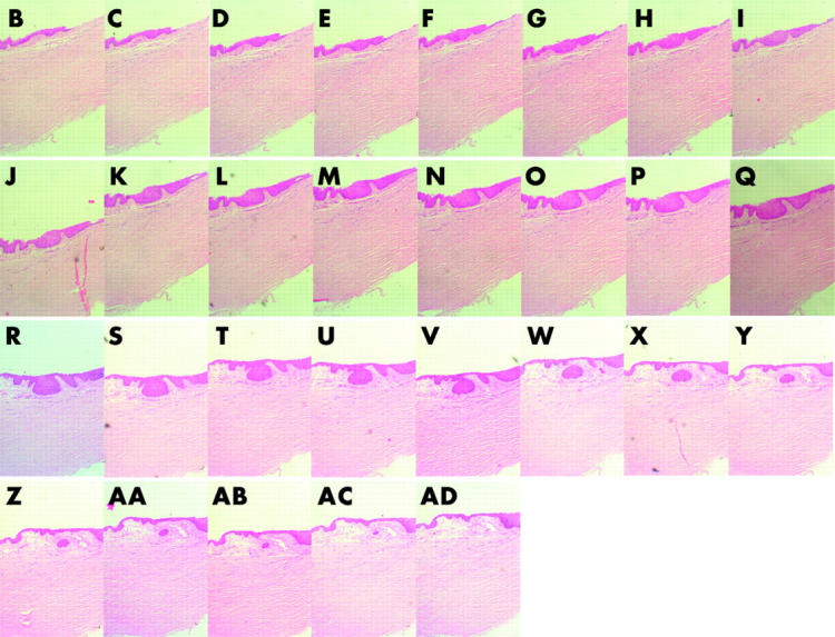
Background/aims: There is substantial evidence that mammalian epithelial stem cells are located within well defined niches. Although the corneoscleral limbus is acknowledged as the site of corneal epithelial stem cells no anatomical niche for such cells has yet been described. The authors undertook to re-evaluate the microanatomy of the limbus in order to identify possible sites that may represent a stem cell niche.
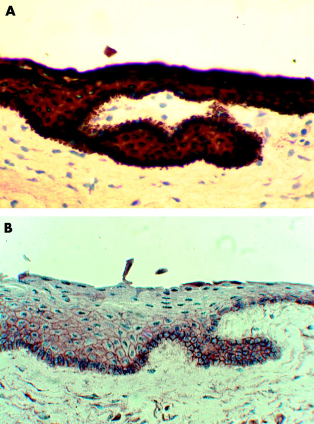
Methods: Systematic serial 5-7 microm sections of human corneoscleral segments obtained from cadaver donors, were examined. The sections were stained with haematoxylin and eosin or toludine blue. Sections with specific areas of interest were further examined immunohistologically for the corneal epithelial marker cytokeratin 14 and the "stem cell" marker ABCG2 transporter protein.
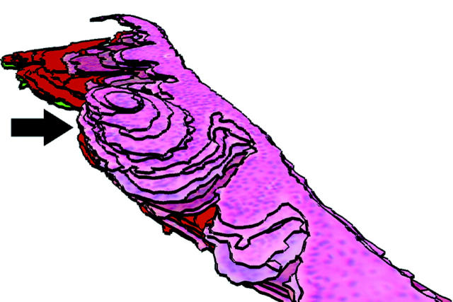
Results: Distinct anatomical extensions from the peripheral aspect of the limbal palisades were identified. These consist of a solid cord of cells extending peripherally or circumferentially. The cells stained positive for CK14 and ABCG2.
Conclusions: A novel anatomical structure has been identified at the human limbus, which demonstrates characteristics of being a stem cell niche. The authors have termed this structure the limbal epithelial crypt.
Figures
References
-
- Fuchs E, Segre J. Stem cells: a new lease of life. Cell 2000;100:143–55. - PubMed
-
- Poulsom R, Alison MR, Forbes SJ, et al. Adult stem cell plasticity. J Pathol 2002;197:441–56. - PubMed
-
- Dua HS, Azuara-Blanco A. Limbal stem cells of the corneal epithelium. Surv Ophthalmol 2000;44:415–425. - PubMed
-
- Dua HS, Forrester JV. Clinical patterns of corneal epithelial wound healing. Am J Ophthalmol 1987;104:481–9. - PubMed
Publication types
MeSH terms
Substances
LinkOut - more resources
Full Text Sources
Medical
Miscellaneous