

Optical coherence tomography characterisation of idiopathic central serous chorioretinopathy
- PMID: 15834085
- PMCID: PMC1772614
- DOI: 10.1136/bjo.2004.049403
Optical coherence tomography characterisation of idiopathic central serous chorioretinopathy
Abstract
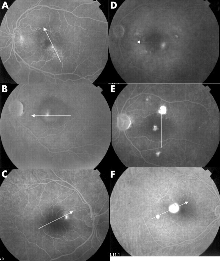
Aim: To describe retinal findings in patients with idiopathic central serous chorioretinopathy (ICSC) as assessed by optical coherence tomography (OCT), and to compare them to fluorescein angiography (FA) findings.
Methods: Case series of 39 eyes from 36 patients with ICSC. Complete ophthalmological examination, last generation OCT (StratusOCT, Software version 3.1) and FA were performed. Six radial scans using OCT were performed and repeated. Singular findings were recorded, OCT images were measured and the results compared with those of FA. The main outcome measures were FA and OCT findings.
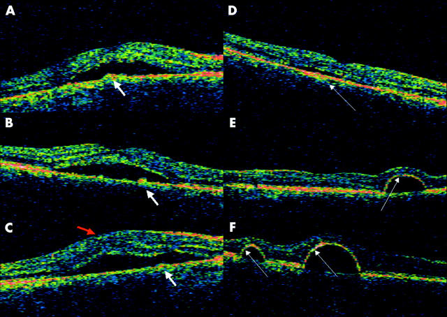
Results: Two patterns of distinct OCT findings are described. In the first one, an optically empty vaulted area of different heights was observed under the neurosensory retina in 36 eyes, being related to fluorescein filled areas; in 35 of them, highly characteristic small bulges could be observed protruding from the retinal pigment epithelium (RPE), angiographically related to leaking spots. In the second pattern, three eyes showed an almost semicircular space under the RPE, with thinner overlying retina.
Conclusions: OCT may offer a new approach to the staging and knowledge of ICSC, and may help the understanding of the mechanisms of the disease.
Figures
References
-
- Gass JDM. Specific diseases causing disciform macular detachment. In: Stereoscopic atlas of macular diseases: diagnosis and treatment. St Louis, Mosby 1997:49–70.
-
- He SZ, Wang W, Li XL, et al. Optical coherence tomography features of central exudative chorioretinopathy. Zhonghua Yan Ke Za Zhi 2003;39:669–72. - PubMed
-
- Cardillo Piccolino F, Eandi CM, Ventre L, et al. Photodynamic therapy for chronic central serous chorioretinopathy. Retina 2003;23:752–63. - PubMed
Publication types
MeSH terms
LinkOut - more resources
Full Text Sources
Medical