

Optic radiation changes after optic neuritis detected by tractography-based group mapping
- PMID: 15834863
- PMCID: PMC6871681
- DOI: 10.1002/hbm.20101
Optic radiation changes after optic neuritis detected by tractography-based group mapping
Abstract
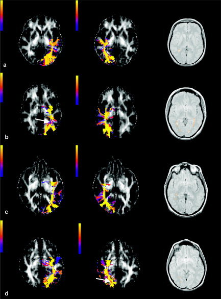
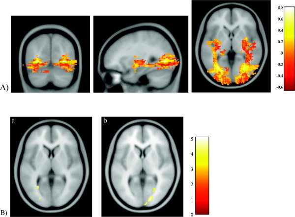
Postmortem data suggest that trans-synaptic degeneration occurs in the lateral geniculate nucleus after optic nerve injury. This study investigated in vivo the optic radiations in patients affected by optic neuritis using fast marching tractography (FMT), a diffusion magnetic resonance imaging (MRI) fiber tracking method, and group mapping techniques, which allow statistical comparisons between subjects. Seven patients, 1 year after isolated unilateral optic neuritis, and ten age and gender-matched controls underwent whole-brain diffusion tensor MR imaging. The FMT algorithm was used to generate voxel-scale connectivity (VSC) maps in the optic radiations in each subject in native space. Group maps of the left and right optic radiations were created in the patient and control group in a standardized reference frame using statistical parametric mapping (SPM99). The reconstructed optic radiations in the patient group were localized more laterally in the posterior part of the tracts and more inferiorly than in the control group. Patients showed reduced VSC values in both tracts compared with controls. These findings suggest that the group mapping techniques might be used to assess changes in the optic radiations in patients after an episode of optic neuritis. The changes we have observed may be secondary to the optic nerve damage.
Figures
References
-
- Ashburner J, Friston KJ (2000): Voxel‐based morphometry—the methods. Neuroimage 11: 805–821. - PubMed
-
- Basser PJ, Mattiello J, LeBihan D (1994): Estimation of the effective self‐diffusion tensor from the NMR spin echo. J Magn Reson B 103: 247–254. - PubMed
-
- Basser PJ, Pajevic S, Pierpaoli C, Duda J, Aldroubi A (2000): In vivo fiber tractography using DT‐MRI data. Magn Reson Med 44: 625–632. - PubMed
-
- Basser PJ, Pierpaoli C (1996): Microstructural and physiological features of tissues elucidated by quantitative‐diffusion‐tensor MRI. J Magn Reson B 111: 209–219. - PubMed
-
- Brusa A, Jones SJ, Plant GT (2001): Long‐term remyelination after optic neuritis: A 2‐year visual evoked potential and psychophysical serial study. Brain 124: 468–479. - PubMed
Publication types
MeSH terms
Grants and funding
LinkOut - more resources
Full Text Sources
Medical
