. 2005 Jun;20(3):131-40.
doi: 10.1111/j.1399-302X.2004.00190.x.

Association of a novel high molecular weight, serine-rich protein (SrpA) with fibril-mediated adhesion of the oral biofilm bacterium Streptococcus cristatus

Affiliations

Association of a novel high molecular weight, serine-rich protein (SrpA) with fibril-mediated adhesion of the oral biofilm bacterium Streptococcus cristatus

P S Handley et al. Oral Microbiol Immunol. 2005 Jun.

Abstract

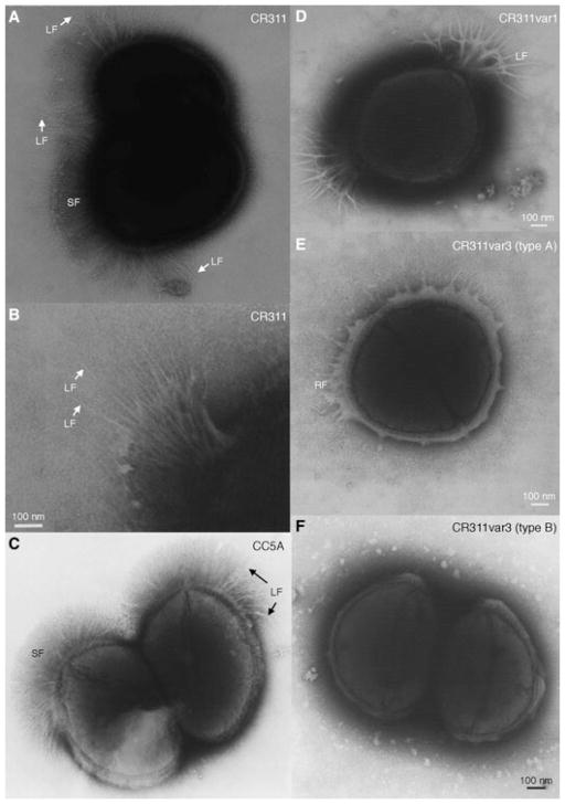
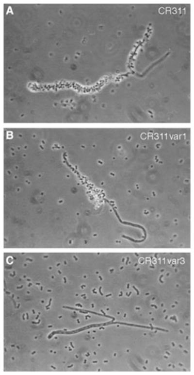
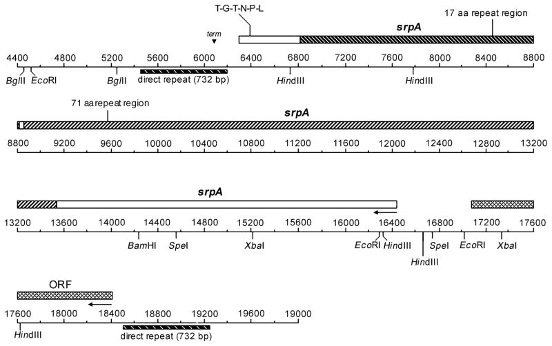
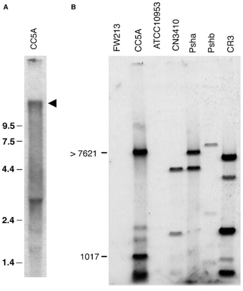
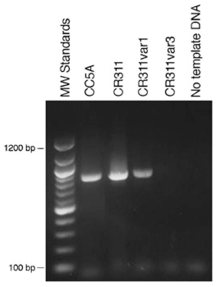
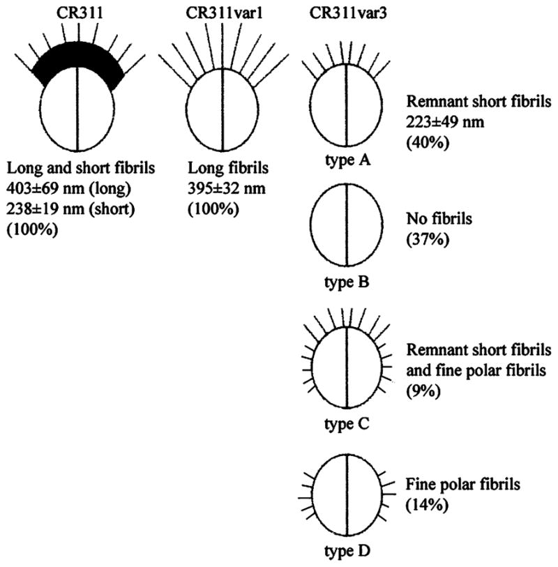
The surface of the oral plaque bacterium Streptococcus cristatus is decorated with a lateral tuft of fibrils. The fibrillar tuft functions in the adhesion of S. cristatus to heterologous bacterial species in the plaque biofilm. The tuft typically consists of a densely packed fringe of shorter fibrils 238 +/- 19 nm long with longer, less abundant fibrils 403 +/- 66 nm long projecting through the fringe of short fibrils. The two types of fibrils in the tufts of S. cristatus have been refractory to biochemical separation, complicating their characterization. A hexadecane partition assay was used to enrich for subpopulations of S. cristatus CR311 (type strain NCTC 12479) having distinct fibrillar morphotypes. Negative staining in the TEM revealed that cells of a hydrophobic subpopulation of S. cristatus (CR311var1) carried only the long fibrils (395 +/- 32 nm). A hydrophilic subpopulation of S. cristatus (CR311var3) consisted of mixed morphotypes having no fibrils or remnant short fibrils (223 +/- 49 nm). No long fibrils were observed on any cells in the CR311var3 subpopulation. The CR311var3 morphotype, unlike the wild-type strain and CR311var1, was not able to form corncobs with either Corynebacterium matruchotii or Fusobacterium nucleatum. Variant CR311var3 did not express the novel gene srpA, which encodes a high molecular weight (321,882 Da) serine-rich protein, SrpA. The SrpA protein contains two extensive repeat motifs of 17 and 71 amino acids and a gram-positive cell wall anchor consensus sequence (LPNTG). The unusual properties of SrpA most closely resemble those of Fap1, the fimbrial-associated adhesin protein of Streptococcus parasanguis. The association of long fibrils, high surface hydrophobicity, ability to form corncob formations, and expression of the srpA gene suggest that SrpA is a long fibril protein in S. cristatus.


Figures

Fig. 1
Fig. 1
Negatively stained preparations of wild-type and fibrillar tuft variants of S. cristatus. A) Wild-type CR311 showing the organization of the short (SF) and long (LF) fibrils. B) Enlargement of the long fibrils in the fibrillar tufts of CR311. C) Wild-type CC5A showing both short and long fibrils. D) CR311var1 showing only the long fibrils that splay out at the ends. E) CR311var3 (type A) showing the remnant tuft fibrils (RF) only. F) CR311var3 (type B) having no fibrils. Magnification bar = 100 nm.
Fig. 2
Fig. 2
Phase contrast microscopy of corncob formation between S. cristatus fibril variants and C. matruchotii. A) Wild-type CR311. B) CR311var1. C) CR311var3. Magnification × 100.
Fig. 3
Fig. 3
Physical map of the region of the S. cristatus CC5A genome containing the srpA ORF. The map shows 14.6 kb of DNA immediately downstream from the binding-deficient locus of S. cristatus CC5A-B15 sequenced previously (5). Two ORFs are depicted by filled bars above the scale. The ORFs reside within a region bounded by a 732 bp direct repeat. Arrows show the direction of transcription. The scale is in bp and numbering continues from the upstream sequence previously published. The complete contiguous 19,841 bp sequence is in the GenBank file (Accession Number U96166).
Fig. 4
Fig. 4
Genetic analysis of the srpA ORF. A) Northern blot showing the srpA transcript from S. cristatus CC5A. Total RNA from wild-type CC5A was hybridized with a DNA sequence amplified by PCR from the 5′-end of the srpA gene. Molecular size standards are shown in kb. The arrow marks the position of a 11 kb hybridization positive band. B) Southern blot of the distribution of srpA DNA in various Streptococcus strains. Total genomic DNA was obtained from S. cristatus strains CC5A, CR3, Psha and Pshb; S. oralis CN3410; S. parasanguis FW213; and F. nucleatum ATCC 10953. The DNA samples were digested to completion with HindIII and the fragments hybridized with a fragment of DNA amplified from the 5′-end of the CC5A srpA ORF. The positions of molecular size markers are shown in bp.
Fig. 5
Fig. 5
Schematic representation of the SrpA protein from S. cristatus CC5A. Organization of the predominant features of the protein are shown. Theoretical glycosylation and phosphorylation sites were determined by computer analysis. Amino acid sequences of the repeat regions and carboxy terminal features of gram-positive wall-anchored proteins were deduced from the nucleotide sequence deposited in GenBank. Sites of amino acid variation within the repetitive sequences are bound by parentheses. Brackets indicate where deletions occur in some of the repeats.
Fig. 6
Fig. 6
PCR analysis of srpA gene sequences in wild-type and fibril variants of CR311. Total DNA from CC5A, CR311, CR311var1 and CR311var3 was amplified in PCR using primers made against the 5′-end of the srpA gene. PCR products were separated on an agarose gel and stained with ethidium bromide. A 100 bp DNA ladder was used as molecular size markers.
Fig. 7
Fig. 7
SDS-polyacrylamide gel analysis of high molecular weight proteins in wild-type and fibril variants of CR311. Proteins from whole cell lysates of CC5A, CR311 and CR311var3 were separated on an 8% SDS-polyacrylamide gel and detected by staining with Coomassie Blue. Molecular weight markers are in kDa. The arrow marks the position of the putative SrpA protein.
Fig. 8
Fig. 8
Population distribution of the various S. cristatus CR311 fibrillar tuft morphological variants. Percent values represent the relative distribution of each fibril type in stationary phase cultures.

References

    1. Brendel V, Trifonov EN. A computer algorithm for testing potential prokaryotic terminators. Nucl Acids Res. 1984;12:4411–4427. - PMC - PubMed
    1. Busscher HJ, Handley PS, Rouxhet P, Hesketh GLM, van der Mei HC. The relationship between structural and physicochemical surface properties of tufted Streptococcus sanguis strains. In: Mozes N, Busscher HJ, Handley PS, Busscher HJ, Rouxhet PG, editors. Microbial Cell Surface Analysis, Structural and Physicochemical Methods. Weinheim; Wiley-VCH: 1991. pp. 319–337.
    1. Chen Q, Wu H, Fives-Taylor PM. Construction of a novel transposon mutagenesis system useful in the isolation of Streptococcus parasanguis mutants defective in Fap1 glycosylation. Infect Immun. 2002;70:6534–6540. - PMC - PubMed
    1. Correia FF, DiRienzo JM, Lamont RJ, Anderman C, McKay TL, Rosan B. Insertional inactivation of binding determinants of Streptococcus crista CC5A using Tn916. Oral Microbiol Immun. 1995;44:287–291. - PMC - PubMed
    1. Correia FF, Lamont RJ, Bayer ME, Rosan B, DiRienzo JM. Cloning and sequencing of a mutated locus that affects fimbrial tuft organization and corncob formation in Streptococcus crista CC5A. Int J Oral Biol. 1997;22:241–248.

Publication types

LinkOut - more resources