Comparative Study
. 2005;132(3):575-80.
doi: 10.1016/j.neuroscience.2004.12.009.

Deletion of presynaptic adenosine A1 receptors impairs the recovery of synaptic transmission after hypoxia

Affiliations
Comparative Study

Deletion of presynaptic adenosine A1 receptors impairs the recovery of synaptic transmission after hypoxia

E Arrigoni et al. Neuroscience. 2005.

Abstract

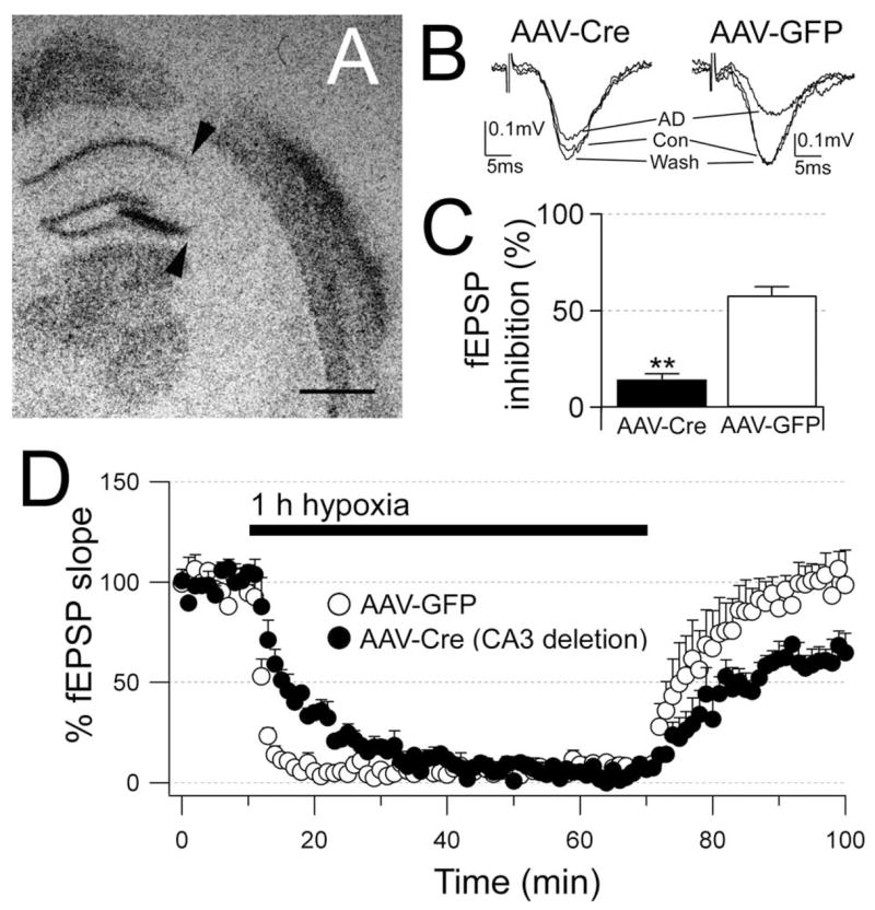
Adenosine protects neurons during hypoxia by inhibiting excitatory synaptic transmission and preventing NMDA receptor activation. Using an adeno-associated viral (AAV) vector containing Cre recombinase, we have focally deleted adenosine A(1) receptors in specific hippocampal regions of adult mice. Recently, we found that deletion of A(1) receptors in the CA1 area blocks the postsynaptic responses to adenosine in CA1 pyramidal neurons, and deletion of A(1) receptors in CA3 neurons abolishes the presynaptic effects of adenosine on the Schaffer collateral input [J Neurosci 23 (2003) 5762]. In the current study, we used this technique to delete A(1) receptors focally from CA3 neurons to investigate whether presynaptic A(1) receptors protect synaptic transmission from hypoxia. We studied the effects of prolonged (1 h) hypoxia on the evoked field excitatory postsynaptic potentials (fEPSPs) in the CA1 region using in vitro slices. Focal deletion of the presynaptic A(1) receptors on the Schaffer collateral input slowed the depression of the fEPSPs in response to hypoxia and impaired the recovery of the fEPSPs after hypoxia. Delayed responses to hypoxia linearly correlated with impaired recovery. These findings provide direct evidence that the neuroprotective role of adenosine during hypoxia depends on the rapid inhibition of synaptic transmission by the activation of presynaptic A(1) receptors.


Figures

Fig. 1
Fig. 1
The A1 receptor antagonist CPT impairs the responses to hypoxia. (A) Electrode positions for the Schaffer collateral stimulations (S) and fEPSPs recording (R). Scale bar=200 μm. (B) Effects of 1 h hypoxia on fEPSPs in the absence (B1) and the presence of 1 μM CPT (B2). Each trace is the average of four consecutive fEPSPs recorded during 1-min periods: just before hypoxia starts (Baseline), after 3 min hypoxia, at the end of the 1 h hypoxia and at the end of the 30 min recovery. (C) Hypoxia quickly and reversibly depresses fEPSP slopes in control conditions (n=4), but in the presence of the A1 receptor antagonist CPT (n=6), hypoxia-induced depression of the fEPSPs occurs at a slower rate and the recovery from hypoxia is impaired. In the graph the fEPSP slopes are represented every 1 min and are expressed as the mean±S.E.M.; 100% is the average of fEPSP slopes during baseline (10 min preceding hypoxia).
Fig. 2
Fig. 2
Focal deletion of the presynaptic A1 receptors impairs the responses to adenosine and hypoxia. (A) In situ hybridization for adenosine A1 receptor mRNA shows deletion of receptors in most of CA3 (arrows). (B) In the same slice, the response of the CA1 fEPSPs to adenosine (50 μM) is markedly reduced (AAV-Cre) compared with the response on the opposite side (AAV-GFP). Each trace is the average of six consecutive fEPSPs. (C) The average response to adenosine in slices with deletion of CA3 A1 receptors (n=4) is significantly reduced compared with the AAV-GFP injection side (n=5). (D) Hypoxia-induced depression of the fEPSPs occurs at a slower rate and the recovery from hypoxia is impaired in slices with focal deletion of the CA3 A1 receptors (n=4) compared with the response in slices from the AAV-GFP injection side (n=5). In the graph the fEPSP slopes are represented every 60 s and are expressed as the mean±S.E.M.; 100% is the average of fEPSP slopes during baseline (10 min preceding hypoxia). Significance by unpaired t-tests P<0.01 (**). Scale bar=1 mm.
Fig. 3
Fig. 3
Incomplete recovery occurs when the response to hypoxia is delayed. (A, B) Application of A1 receptor antagonists and focal deletion of the presynaptic A1 receptors on the Schaffer collateral input delays hypoxia-mediated fEPSP depression and impairs recovery from hypoxia. The two graphs compare the response to hypoxia in slices from wild-type mice in normal ACSF and in the presence of CPT (1 μM) and in inducible A1 receptor knock-out mice in slices from the AAV-GFP-injected side and from the AAV-Cre-injected side. (C) Delay in the onset of the hypoxia-mediated fEPSP depression inversely correlates with lack of full recovery from hypoxia. Summary of all experiments: wild-type mice are represented with triangles with recordings in normal ACSF (open triangles; n=4) and in the presence of CPT (filled triangles; n=6); inducible receptor knock-out mice are A1 represented with circles with the recordings from the AAV-Cre-injected side (filled circles; n=4) and from the AAV-GFP-injected side (open circles; n=5). The onset of the fEPSP depression is determined by best fit with single exponential functions. Values are expressed as mean±S.E.M.; 100% is the average of fEPSP slopes during baseline (10 min preceding hypoxia). Significance by unpaired t-tests P<0.01 (**); P<0.05 (*).

References

    1. Boening JA, Kass IS, Cottrell JE, Chambers G. The effect of blocking sodium influx on anoxic damage in the rat hippocampal slice. Neuroscience. 1989;33:263–268. - PubMed
    1. Coelho JE, de Mendonca A, Ribeiro JA. Presynaptic inhibitory receptors mediate the depression of synaptic transmission upon hypoxia in rat hippocampal slices. Brain Res. 2000;869:158–165. - PubMed
    1. Costenla AR, De Mendonca A, Sebastiao A, Ribeiro JA. An adenosine analogue inhibits NMDA receptor-mediated responses in bipolar cells of the rat retina. Exp Eye Res. 1999;68:367–370. - PubMed
    1. Dale N, Pearson T, Frenguelli BG. Direct measurement of adenosine release during hypoxia in the CA1 region of the rat hippocampal slice. J Physiol. 2000;526(Pt 1):143–155. - PMC - PubMed
    1. de Mendonca A, Ribeiro JA. Contribution of metabotropic glutamate receptors to the depression of excitatory postsynaptic potentials during hypoxia. Neuroreport. 1997;8:3667–3671. - PubMed

Publication types

MeSH terms