. 2005 Apr 25;169(2):257-68.
doi: 10.1083/jcb.200501156. Epub 2005 Apr 18.

Transcriptional regulation of myotube fate specification and intrafusal muscle fiber morphogenesis

Affiliations

Transcriptional regulation of myotube fate specification and intrafusal muscle fiber morphogenesis

Y'vonne Albert et al. J Cell Biol. .

Abstract

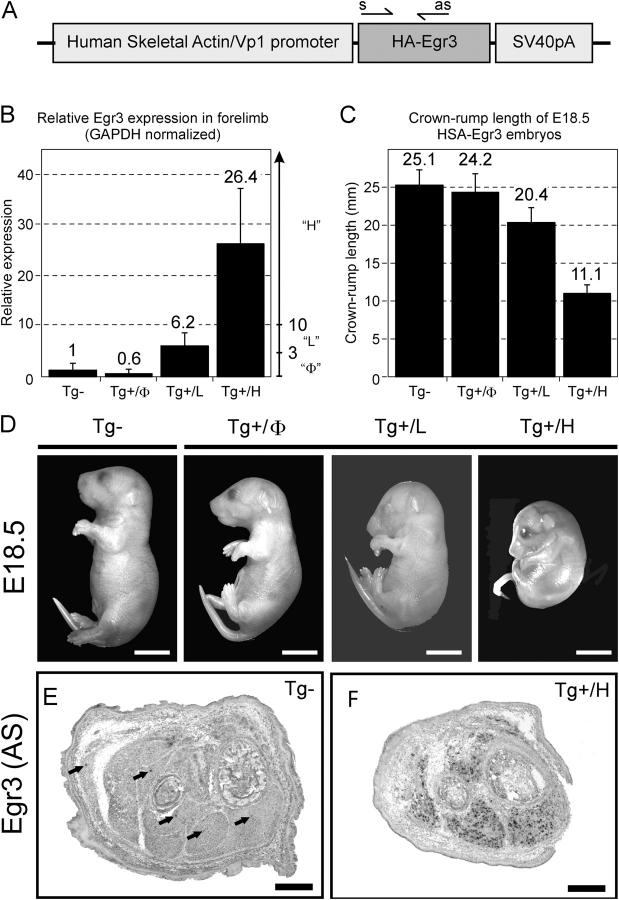
Vertebrate muscle spindle stretch receptors are important for limb position sensation (proprioception) and stretch reflexes. The structurally complex stretch receptor arises from a single myotube, which is transformed into multiple intrafusal muscle fibers by sensory axon-dependent signal transduction that alters gene expression in the contacted myotubes. The sensory-derived signal transduction pathways that specify the fate of myotubes are very poorly understood. The zinc finger transcription factor, early growth response gene 3 (Egr3), is selectively expressed in sensory axon-contacted myotubes, and it is required for normal intrafusal muscle fiber differentiation and spindle development. Here, we show that overexpression of Egr3 in primary myotubes in vitro leads to the expression of a particular repertoire of genes, some of which we demonstrate are also regulated by Egr3 in developing intrafusal muscle fibers within spindles. Thus, our results identify a network of genes that are regulated by Egr3 and are involved in intrafusal muscle fiber differentiation. Moreover, we show that Egr3 mediates myotube fate specification that is induced by sensory innervation because skeletal myotubes that express Egr3 independent of other sensory axon regulation are transformed into muscle fibers with structural and molecular similarities to intrafusal muscle fibers. Hence, Egr3 is a target gene that is regulated by sensory innervation and that mediates gene expression involved in myotube fate specification and intrafusal muscle fiber morphogenesis.


Figures

Figure 1.
Figure 1.
Identification of Egr3 target genes in primary murine myotubes. (A) Adenoviruses were generated to express either EGFP alone (EGFP), full-length, transcriptionally active Egr3 and EGFP (Egr3WT), or transcriptionally inactive Egr3 and EGFP (Egr3Tr). The transcriptional activity of the recombinant proteins that were produced by the viruses was assessed by the early growth response element (ERE) luciferase reporter assay. Compared with EGFP and Egr3Tr-infected myoblasts, Egr3WT infection showed marked transcriptional activation of the ERE–luciferase reporter plasmid (mean and standard deviation shown from two independent transfection-infection experiments, each performed in triplicate). (B) Myotubes were differentiated for 10 d and infected with either the Egr3WT or Egr3Tr adenovirus. EGFP fluorescence confirmed 100% infection efficiency, and immunohistochemistry using a COOH-terminal Egr3 antibody demonstrated high levels of nuclear localized Egr3 in Egr3WT-infected myotubes and no staining in the Egr3Tr-infected myotubes. Results from the Egr3Tr-infected cells further demonstrated the lack of endogenous Egr3 expression in wild-type myotubes. Bar, 40 μm. (C) Total RNA was extracted from the myotubes 18 h after infection, and Affymetrix microarray analysis was performed. The infection experiments were performed twice, and the gene expression datasets were compared in a four-way analysis. 83 genes were identified as up-regulated in all of the single, four-way comparisons from 45,200 genes and transcripts that were analyzed. (D) All of the genes tested (15/15) were confirmed to represent up-regulated genes by real-time PCR (mean and standard deviation from four independent infection experiments and real-time PCR assays, each performed in triplicate, P < 0.05 for all genes).
Figure 2.
Figure 2.
Pea3 expression in wild-type and Egr3 -deficient muscle is a marker of Ia-afferent–contacted myotubes in vivo. (A) At E16.5, shortly after Ia-afferent contact is made with myotubes, Pea3 is expressed in both wild-type and Egr3-deficient myotubes. In Egr3-deficient muscles, the number of myotubes that express Pea3 is reduced by 66% in E16.5 embryos, and (B) in E18.5 embryos, they are reduced by 86%. Ia-afferents are known to initially contact myotubes in Egr3-deficient mice and then withdraw during development, which is consistent with the progressive decrease in Pea3-expressing myotubes that are identified in Egr3-deficient muscles. Thus, Pea3 is not regulated by Egr3 in myotubes, and it is a marker for Ia-afferent contact. These results make it possible to use Pea3 to localize Ia-afferent–contacted myotubes and study potential Egr3 target gene expression within them (n = 3 animals for each genotype; mean and standard deviation shown). Insets are high power magnifications of labeled spindles (arrows). Bars: 50 μm; (inset) 10 μm.
Figure 3.
Figure 3.
Egr3 regulates specific target genes in developing spindles. Affymetrix microarray analysis demonstrated many potential target genes that are regulated by Egr3 in myotubes. Egr3 was necessary for the expression of (A) NGFR (p75), (B) SSTr2, and (C) Prph1 in muscle spindles. The muscle expression of all of these genes was restricted to the developing spindles (arrows), similar to the expression pattern of Egr3, whereas their expression was abrogated in Pea3-expressing (Ia-afferent contacted), Egr3-deficient myotubes. Bars: 50 μm; (inset) 10 μm.
Figure 4.
Figure 4.
Enforced Egr3 expression in all skeletal myotubes leads to muscle defects and perinatal mortality. (A) The human skeletal actin (HSA) promoter was used to express Egr3 in all skeletal myotubes independent of normal Ia-afferent–mediated regulation. PCR was used to genotype the mice using the primers indicated (s and as). (B) Perinatal founder mice were not viable, making it necessary to characterize all of the individual embryos generated from the pronuclear injections. Using real-time PCR to examine the expression of Egr3 in muscle, a total of 58 embryos were studied by separating them into four groups: (1) those that did not carry the transgene (Tg−); (2) those that carried the transgene but did not express it (Tg+/Φ); (3) those that carried the transgene and expressed it at low levels (Tg+/L; 3–10-fold increase in Egr3 expression relative to Tg− muscles); and (4) those that carried the transgene and expressed it at high levels (Tg+/H; >10-fold increase in Egr3 expression relative to Tg− muscles; n = 8–20 embryos per group; mean and standard deviation shown). (C) The transgene-expressing mice were consistently smaller than either their Tg− or Tg+/Φ litter mates (n = 8–20 embryos per group; mean and standard deviation shown). (D) The late gestational (E18.5) Tg+/L and Tg+/H embryos had beating hearts, flexion contractions, and exhibited no spontaneous or evoked motor activity. Bars, 5 mm. (E) In situ hybridization demonstrated normal Egr3 expression in developing spindles in both Tg− and Tg+/Φ muscles (arrows). (F) In contrast, Tg+ mice expressed the transgene in most muscle fibers, many of which were aggregated into fibers with numerous internal nuclei. Bars, 0.5 mm.
Figure 5.
Figure 5.
Skeletal myotubes are transformed into muscle fibers that are structurally similar to intrafusal muscle fibers in HSA– Egr3 transgenic mice. E18.5 (A, C, and E) wild-type and (B, D, and F) Tg+/H transgenic hindlimbs. (A) In wild-type embryos, skeletal muscles were discretely formed (lower hindlimb shown), whereas (B) in HSA–Egr3 transgene-expressing embryos, the individual muscles were poorly distinguished. Boxed areas in A and B are shown in C and D at higher magnifications. (C and E) Discrete wild-type muscles contained normal muscle fibers that were distended by skeletal myofilaments and contained subsarcolemmal nuclei. (C) Occasional normal spindles were noted (arrowhead). (E) They contained three to four intrafusal muscle fibers with scant myofibrillary structure and internal nuclei, which were surrounded by axons (Ia-afferents) and rudimentary capsules (arrowhead). (D) Muscle fibers from transgene-expressing mice had scant myofibrillary structure and internal nuclei similar to those of intrafusal muscle fibers. (F) Many of the fibers were aggregated into spindle-like structures that lacked capsules or obvious innervation (arrow). Scattered, normal-appearing spindles were noted to have capsules and innervation (arrowhead). Bars: (A and B) 500 μm; (C and D) 20 μm; (E and F) 100 μm.
Figure 6.
Figure 6.
Intrafusal-like muscle fibers are not innervated in HSA– Egr3 transgene-expressing mice. (A) A subset of proprioceptive neurons in the dorsal root ganglia (DRG) give rise to Ia-afferents that express parvalbumin (Pv). There was no difference in the number of Pv+ neurons in DRG between Tg− and Tg+/H mice (fifth lumbar DRG shown), which is consistent with normal innervation of some spindles in Tg+/H muscles (n = 4 for each genotype; mean and standard deviation shown). (B) However, although Pv+ Ia-afferents were robustly labeled in normal spindles in Tg− (arrowhead) and Tg+/H mice (not depicted), there was no evidence of Pv+ axon innervation in any of the transformed myotubes in Tg+/H muscles. (C) Muscles in Tg+/H mice were not capable of sustaining any motor innervation. Motor neurons (mn) present in Tg− spinal cords (lumbar cord shown) were entirely absent in Tg+/H mice, leaving only spinal interneurons in their place. Complete motor neuron loss was corroborated by the absence of ventral roots (vr), which carry motor axons into the periphery. (D) Spindles in Tg− muscles received both Ia-afferent and fusimotor innervation, which was robustly labeled by ATP1α3 immunohistochemistry, whereas no ATP1α3-labeled axons innervated the transformed myotubes in Tg+/H muscles. Arrowheads indicate the spindles innervated by ATPα3-containing axons. (E) Consistent with the observation that Pv+ neurons are not altered in Tg+/H mice, Ia-afferent–contacted, Pea3-expressing myotubes were observed in Tg+/H muscles, and Pea3 expression was not significantly different from wild-type (Tg−) or nontransgene-expressing (Tg+/Φ) muscles (n = 4 for each condition; mean and standard deviation shown). Inset shows a magnified image of the Pea3-expressing spindle (arrow). Bars: (A, C, and E) 50 μm; (B and D) 20 μm; (inset) 10 μm.
Figure 7.
Figure 7.
Muscle fibers in HSA– Egr3 transgenic mice express genes that are characteristic of intrafusal muscle fibers. (A) Slow developmental myosin heavy chain (Sd-MyHC), a well-established marker for intrafusal muscle fibers, was expressed selectively by the relatively few intrafusal muscle fibers present in wild-type muscles (arrows), whereas it was markedly up-regulated by muscle fibers in Tg+/H mice. (bar graph) The number of Sd-MyHC–expressing myotubes was significantly increased in Tg+ relative to Tg− mice when the number of myotubes/section were counted in lower hindlimb muscles. Bars: 50 μm; (inset) 25 μm. (B) Similarly, expression of the neurotrophins NT-3 and GDNF, both of which are expressed in muscle primarily by intrafusal muscle fibers in E18.5 wild-type mice, were up-regulated in Tg+/H muscles but not in nontransgene-expressing muscles (Tg+/Φ) relative to wild type (Tg−). (C) The expression of NGFR (p75), SSTr2, and Prph1, three newly identified target genes that are regulated by Egr3 in spindles, were also markedly up-regulated, as were (D) several other genes (ATP1α3, Hey1, and 2810417MoRik) that were examined from the Egr3 target gene microarray analysis results (mean and standard deviation shown; n = 4 embryos for each genotype; *, P < 0.05; **, P < 0.01).

Similar articles

Cited by

References

    1. Andrechek, E.R., W.R. Hardy, A.A. Girgis-Gabardo, R.L. Perry, R. Butler, F.L. Graham, R.C. Kahn, M.A. Rudnicki, and W.J. Muller. 2002. ErbB2 is required for muscle spindle and myoblast cell survival. Mol. Cell. Biol. 22:4714–4722. - PMC - PubMed
    1. Arber, S., D.R. Ladle, J.H. Lin, E. Frank, and T.M. Jessell. 2000. ETS gene Er81 controls the formation of functional connections between group Ia sensory afferents and motor neurons. Cell. 101:485–498. - PubMed
    1. Chen, H.H., W.G. Tourtellotte, and E. Frank. 2002. Muscle spindle-derived neurotrophin 3 regulates synaptic connectivity between muscle sensory and motor neurons. J. Neurosci. 22:3512–3519. - PMC - PubMed
    1. Chen, Y.W., P. Zhao, R. Borup, and E.P. Hoffman. 2000. Expression profiling in the muscular dystrophies: identification of novel aspects of molecular pathophysiology. J. Cell Biol. 151:1321–1336. - PMC - PubMed
    1. Crawford, G.E., J.A. Faulkner, R.H. Crosbie, K.P. Campbell, S.C. Froehner, and J.S. Chamberlain. 2000. Assembly of the dystrophin-associated protein complex does not require the dystrophin COOH-terminal domain. J. Cell Biol. 150:1399–1410. - PMC - PubMed

Publication types

MeSH terms