. 2005 Apr 18;201(8):1197-203.
doi: 10.1084/jem.20050158.

IL-7 receptor signaling is necessary for stage transition in adult B cell development through up-regulation of EBF

Affiliations

IL-7 receptor signaling is necessary for stage transition in adult B cell development through up-regulation of EBF

Kazu Kikuchi et al. J Exp Med. .

Abstract

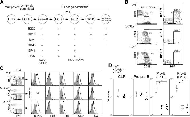
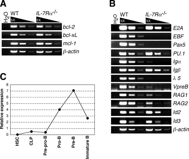
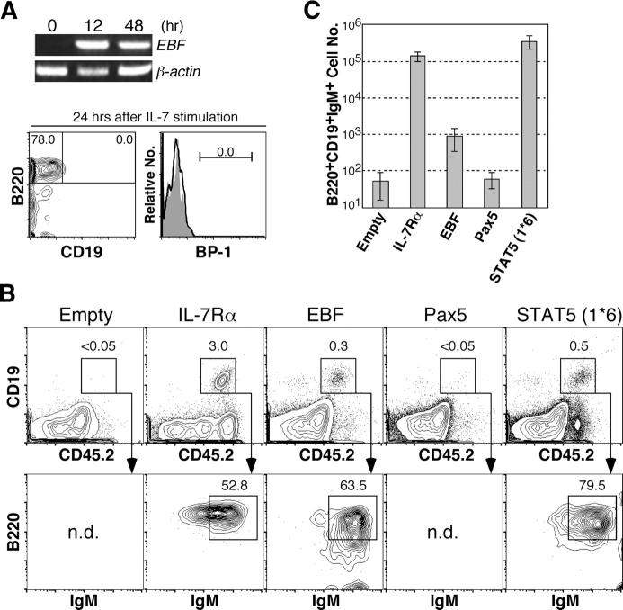
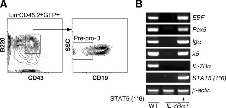
Cytokine receptor signals have been suggested to stimulate cell differentiation during hemato/lymphopoiesis. Such action, however, has not been clearly demonstrated. Here, we show that adult B cell development in IL-7(-/-) and IL-7R alpha(2/-) mice is arrested at the pre-pro-B cell stage due to insufficient expression of the B cell-specific transcription factor EBF and its target genes, which form a transcription factor network in determining B lineage specification. EBF expression is restored in IL-7(-/-) pre-pro-B cells upon IL-7 stimulation or in IL-7R alpha(-/-) pre-pro-B cells by activation of STAT5, a major signaling molecule downstream of the IL-7R signaling pathway. Furthermore, enforced EBF expression partially rescues B cell development in IL-7R alpha(-/-) mice. Thus, IL-7 receptor signaling is a participant in the formation of the transcription factor network during B lymphopoiesis by up-regulating EBF, allowing stage transition from the pre-pro-B to further maturational stages.


Figures

Figure 1.
Figure 1.
B cell development is arrested at the pre–pro-B stage in the absence of IL-7R signaling. (A) Schematic maturational relationship of B cell populations used in this study. We excluded Ly-6C+ and NK1.1+ cells from Hardy's fraction A (reference 25) except for the experiments shown in Fig. 4. CD19 can be used instead of HSA to distinguish pre–pro-B (fraction A) and pro-B (fractions B and C/C′) cells (reference ; Fig. S1 D, available at http://www.jem.org/cgi/content/full/jem.20050158/DC1). Hereafter, we used the fraction C/C′ population as fraction C. (B) Bone marrow (BM) cells from 8-wk-old mice were stained with antibodies for lineage markers (Lin: CD3ɛ, Gr-1, Ter119, and Mac-1), B220, CD43, BP-1, and HSA. Propidium iodide+ dead cells as well as Lin+ cells were excluded from this cell surface phenotyping. The numbers in FACS plots are percentages of cells in each cell population for each plot. The representative results of at least three independent experiments are shown. (C) The expression of IL-7Rα, c-Kit, Flt3, AA4.1, and HSA in pre–pro-B cells derived from WT, IL-7Rα−/−, and IL-7 −/− mice. The definition of pre–pro-B cells here is LinB220+CD43+CD19NK1.1Ly-6C cells. Removal of CD19+ cells from this analysis is critical to exclude the more mature B cell progenitors from the pre–pro-B cell population (see Fig. S1). Open and filled histograms represent the expression level of various markers and the negative control stained with isotype-matched irrelevant antibodies, respectively. We did not stain pre–pro-B cells from IL-7Rα−/− mice with anti–IL-7Rα antibodies in this analysis. (D) The number of CLP, pre–pro-B, and pro-B cells in bone marrow from bilateral femurs and tibiae derived from WT (open circle), IL-7Rα−/− (closed circle), and IL-7 −/− (closed square) mice. The absolute number of cells was calculated with total bone marrow cell numbers and the percentage of each cell population. The mean is indicated as a horizontal bar. Because IL-7Rα is a marker to define CLPs, we did not determine the number of CLPs in IL-7Rα−/− mice. Asterisks denote significant difference by Student's t test; P < 0.05.
Figure 2.
Figure 2.
Gene expression profiles in IL-7R α / pre–pro-B cells. (A) Expression of antiapoptotic genes in WT and IL-7Rα−/− pre–pro-B cells were examined by RT-PCR. RNA was purified from 1.5 × 10⁴ pre–pro-B cells (LinB220+CD43+CD19NK1.1Ly-6C cells), which were sorted twice in sequence (double sorted) from WT and IL-7Rα−/− BM cells. After double sorting, the purity of cells was >99% in reanalysis. (B) Expression of genes that are involved in B cell development in WT and IL-7Rα−/− pre–pro-B cells. RT-PCR analyses were done as in A. (C) Expression of EBF gene in various developing B cell populations. The cell populations used here are HSC (Lin−/lowThy-1.1lowc-KithighSca-I+), CLP (Linc-KitlowSca-IlowThy-1.1IL-7Rα+), pre–pro-B (B220+CD43+CD19NK1.1Ly-6C), pro-B (B220+CD43+CD19+HSAhigh), pre-B (B220+CD43IgM), and immature B (B220+IgM+IgD). The lineage cocktail (Lin) used for HSCs and CLPs also contained anti-B220 antibodies. Total RNA was purified from each doubly sorted population and subjected to quantification of EBF mRNAs by real-time PCR after first strand synthesis with reverse transcriptase. The amount of the first strand DNA applied was normalized to the expression level of a reference gene, GAPDH. EBF expression in whole BM was arbitrarily defined as unit one. The mean value of more than three independent samples is shown. Range of error is too small to be displayed in the histogram.
Figure 3.
Figure 3.
EBF expression is regulated by IL-7R signaling in B cell development. (A) Restoration of EBF expression in IL-7 −/− pre–pro-B cells. 1.2 × 10⁴ pre–pro-B cells (B220+CD43+CD19NK1.1Ly-6C) derived from IL-7 −/− mice were cultured in the presence of 10 ng/ml IL-7 in 96-well plates. The same number of freshly isolated cells was used as a nonstimulated control (time 0). Cells were harvested at the indicated time points and subjected to RT-PCR analysis of EBF expression. We also checked the cell surface phenotype of cells at 24 h after the culture. At this time point, no pro-B cells (CD19+ or BP-1+ cells) were developed from pre–pro-B cells (open histogram). Closed histogram represents background staining with isotype control antibodies. (B) IL-7Rα−/− HSCs (CD45.2+) were infected with empty, IL-7Rα, EBF, Pax5, or STAT5 (*6) retroviruses by spin infection and injected into sublethally irradiated RAG2 −/− mice (CD45.1+). 5 wk after injection, mice were killed and spleen cell suspensions were stained with antibodies for CD19, B220, and IgM, as well as for CD45.2. CD19+B220+IgM+ cells are mature B cells. Representative data from more than three samples are shown. We did not observe any immature B cells derived from EBF+ IL-7Rα−/− HSCs in bone marrow at this time point but did observe them at an earlier time point (Fig. S2, available at http://www.jem.org/cgi/content/full/jem.20050158/DC1). (C) CD19+B220+IgM+ cell numbers of reconstituted mice with IL-7Rα2/− HSCs infected with empty, IL-7Rα, EBF, Pax5, or STAT5 (*6) retroviruses shown in B were calculated. The cell numbers are expressed as mean ± SD.
Figure 4.
Figure 4.
STAT5, a downstream signaling molecule of IL-7R, mediates EBF gene expression. (A) Donor-derived pre–pro-B cells (CD45.2+GFP+B220+CD43+CD19) were purified from reconstituted mice with IL-7Rα−/− HSCs infected with STAT5 (*6) retrovirus. (B) 1.0 × 10⁴ pre–pro-B cells derived from WT (left lane), IL-7Rα−/− mice (middle lane), and reconstituted mice with IL-7Rα−/− HSCs expressing STAT5 (*6) (right lane) were used for analysis of B cell–specific gene expression by RT-PCR. EBF expression as well as expression of its targets, Pax5, Igα, and λ5, were restored in pre–pro-B cells in the presence of constitutive active STAT5. Because IL-7Rα expression was not detected in the right lane, there was no contamination of host-derived cells (RAG2 −/− mice) in the sample.

References

    1. Quong, M.W., W.J. Romanow, and C. Murre. 2002. E protein function in lymphocyte development. Annu. Rev. Immunol. 20:301–322. - PubMed
    1. Busslinger, M. 2004. Transcriptional control of early B cell development. Annu. Rev. Immunol. 22:55–79. - PubMed
    1. Kee, B.L., and C. Murre. 1998. Induction of early B cell factor (EBF) and multiple B lineage genes by the basic helix-loop-helix transcription factor E12. J. Exp. Med. 188:699–713. - PMC - PubMed
    1. Medina, K.L., J.M. Pongubala, K.L. Reddy, D.W. Lancki, R. Dekoter, M. Kieslinger, R. Grosschedl, and H. Singh. 2004. Assembling a gene regulatory network for specification of the B cell fate. Dev. Cell. 7:607–617. - PubMed
    1. O'Riordan, M., and R. Grosschedl. 1999. Coordinate regulation of B cell differentiation by the transcription factors EBF and E2A. Immunity. 11:21–31. - PubMed

Publication types

MeSH terms