. 2005 Apr 18;201(8):1281-92.
doi: 10.1084/jem.20041276.

Neutrophils mediate immune modulation of dendritic cells through glycosylation-dependent interactions between Mac-1 and DC-SIGN

Affiliations

Neutrophils mediate immune modulation of dendritic cells through glycosylation-dependent interactions between Mac-1 and DC-SIGN

Klaas P J M van Gisbergen et al. J Exp Med. .

Abstract

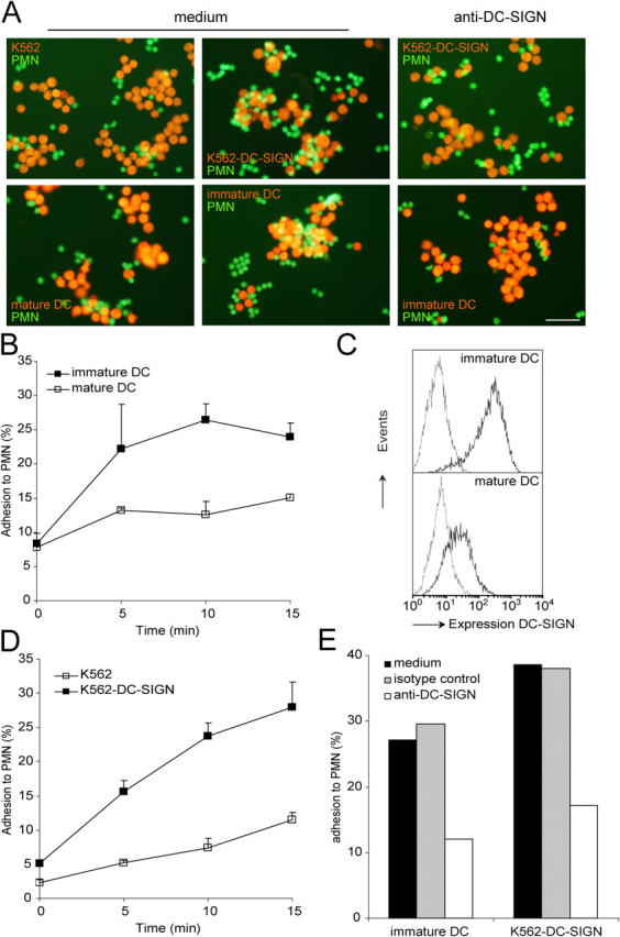
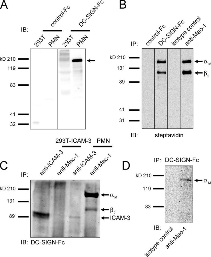
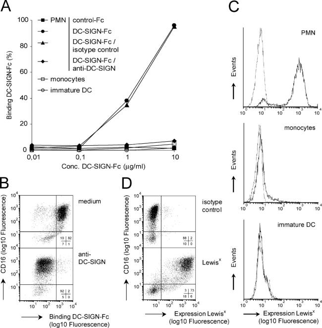
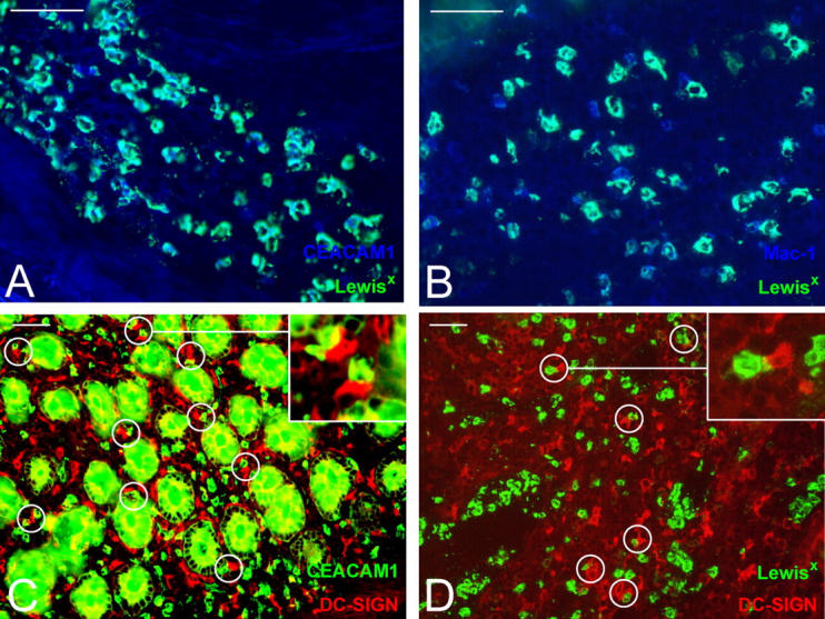
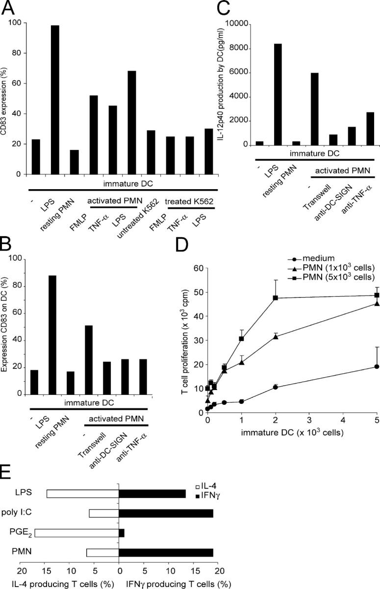
Neutrophils are key players of the innate immune system that provide a first line of defense against invading pathogens. However, it is unknown whether neutrophils can interact with dendritic cells (DCs) to modulate adaptive immune responses. We demonstrate that neutrophils strongly cluster with immature DCs and that activated, not resting, neutrophils induce maturation of DCs that enables these DCs to trigger strong T cell proliferation and T helper type 1 polarization of T cells. This neutrophil-DC interaction is driven by the binding of the DC-specific, C-type lectin DC-SIGN to the beta(2)-integrin Mac-1. Strikingly, DC-SIGN only interacts with Mac-1 from neutrophils, but not from other leukocytes, mainly because of specific Lewis(x) carbohydrates that are present on the alpha(M) chain of Mac-1 from neutrophils. Furthermore, we show that besides the formation of cellular contact, the tumor necrosis factor-alpha produced by activated neutrophils is essential for inducing DC maturation. Our data demonstrate that DC-SIGN and Mac-1 define a molecular pathway to establish cellular adhesion between DCs and neutrophils, thereby providing a novel cellular link between innate and adaptive immunity.


Figures

Figure 1.
Figure 1.
DC-SIGN mediates clustering of DCs and PMN. (A) CFSE-labeled PMN (green) were incubated with hydroethidine-labeled K562, K562-DC-SIGN, immature DCs, or mature DCs (red) for 30 min at 37°C. Anti–DC-SIGN antibodies (AZN-D1 and AZN-D2; 20 μg/ml) were used to block DC-SIGN dependent adhesion. Cell–cell clustering was visualized using fluorescent microscopy and representative pictures were taken. Bar, 50 μm. Three independent experiments with similar results were performed. (B) Using FACS analysis cell–cell clustering was followed in time by scoring the percentage of immature or mature DCs that have bound PMN. (C) The expression of DC-SIGN was analyzed on immature and mature DCs by flow cytometry. (D) The percentage of K562 or K562-DC-SIGN that have bound PMN was measured in time by FACS analysis. (E) DC-SIGN–dependent adhesion of PMN to immature DCs and K562-DC-SIGN was determined with blocking anti–DC-SIGN antibodies at 10 min of cell–cell clustering. One out of three independent experiments with similar results is shown.
Figure 2.
Figure 2.
Identification of PMN Mac-1 as a novel ligand of DC-SIGN. (A) Whole PMN and 293T lysate were analyzed on immunoblot (IB) with control-Fc and DC-SIGN-Fc. The arrow indicates the main DC-SIGN ligand of ∼160 kD. (B) Surface-biotinylated PMN were lysed and immunoprecipitated (IP) with control-Fc, DC-SIGN-Fc, mouse IgG1 isotype control antibodies, and anti–Mac-1 (αM chain) antibodies. Immunoprecipitates were analyzed on immunoblot with streptavidin. Arrows indicate the αM and β2 chains of Mac-1. (C) 293T-ICAM-3 and PMN were immunoprecipitated with anti–Mac-1 and anti–ICAM-3 antibodies and immunoblotted with DC-SIGN-Fc. Arrows indicate ICAM-3 and the αM and β2 chains of Mac-1. (D) DC-SIGN-Fc immunoprecipitates of PMN lysate were immunoblotted with isotype control and anti–Mac-1 (αM chain) antibodies. Arrow indicates the αM chain of Mac-1. Results are representative of three independent experiments.
Figure 3.
Figure 3.
DC-SIGN specifically binds Lewisx-expressing PMN. (A) The binding of control-Fc and DC-SIGN-Fc to PMN, monocytes, and immature DCs was analyzed by flow cytometry. Binding of DC-SIGN-Fc to PMN is sensitive to blocking anti–DC-SIGN antibodies (AZN-D1; 50 μg/ml). (B) DC-SIGN-Fc preferably binds CD16+ neutrophils and not CD16 eosinophils (top). Blocking anti–DC-SIGN antibodies (AZN-D1; 50 μg/ml) inhibit binding of DC-SIGN-Fc (bottom). Insets represent the percentage of cells within a quadrant. (C) Isotype (thin line) and Lewisx (thick line) expressions were measured on PMN, monocytes, and immature DCs. (D) Isotype (top) and Lewisx expression (bottom) were examined on CD16+ neutrophils and CD16 eosinophils. Insets represent the percentage of cells within a quadrant. One out of three independent experiments with similar results is shown.
Figure 4.
Figure 4.
DC-SIGN binds Mac-1 expressed on PMN through Lewisx carbohydrates. (A) Sandwich ELISA on ICAM-3, LFA-1, Mac-1, p150,95, and β2-integrins from PMN. (B) The binding of DC-SIGN-Fc to ICAM-3 that was captured from mock and ICAM-3–transfected 293T cells was measured in an ELISA-based binding assay. (C) DC-SIGN-Fc binding to ICAM-3, LFA-1, Mac-1, p150,95, and β2-integrins that were captured from PMN (white bars) and the reverse reaction—the capture of DC-SIGN ligands using DC-SIGN-Fc and the detection of these ligands using antibodies (black bars)—were measured in an ELISA-based binding assay. (D) Using ELISA, the presence of Lewisx on LFA-1, Mac-1, p150,95, and β2-integrins derived from PMN was determined. (E) The quantity of Mac-1 of PMN, monocytes, and immature DCs was determined using a sandwich ELISA for Mac-1 (capture, αM chain; detection, β2 chain). (F) DC-SIGN-Fc binding to Mac-1 captured from lysate of PMN, monocytes, and immature DCs was analyzed in an ELISA-based binding assay. Ca2+ specificity of DC-SIGN binding was determined using the Ca2+ chelator EGTA (10 mM). (G) The presence of Lewisx on Mac-1 that was derived from PMN, monocytes, and immature DCs was measured by ELISA (capture, Mac-1; detection, Lewisx). (H) Lysates of surface-biotinylated PMN were immunoprecipitated with anti–Mac-1 antibodies. The immunoprecipitates were incubated overnight with or without α1-3,4-fucosidase (Xanthomonas; Calbiochem) in sodium phosphate buffer (50 mM, pH 5) at 37°C. Immunoprecipitates were then immunoblotted with streptavidin, anti-Lewisx antibodies, and DC-SIGN-Fc. Arrows indicate the αM and β2 chains of Mac-1. Results are representative of three independent experiments.
Figure 5.
Figure 5.
The C-type lectin domain of DC-SIGN binds PMN Mac-1. (A) The binding of fluorescent beads coated with PMN-derived LFA-1, p150,95, and Mac-1 was measured to K562-DC-SIGN. Adhesion of PMN-derived Mac-1 beads was sensitive to blocking anti–DC-SIGN antibodies (AZN-D1; 20 μg/ml). (B) The adhesion of Mac-1 derived from PMN, monocytes, and immature DCs to K562-DC-SIGN was analyzed in a fluorescent bead adhesion assay. Anti–DC-SIGN antibodies were used to determine the specificity of DC-SIGN binding. (C) The adhesion of fluorescent beads coated with PMN Mac-1 to K562 cells and K562 transfectants expressing wildtype or mutant DC-SIGN, in which amino acids within the C-type lectin domain essential for ligand binding or the positioning of Ca2+ ions are mutated, is shown. One out of three independent experiments is shown.
Figure 6.
Figure 6.
In vivo localization of PMN and DCs in colonic mucosa of patients with Crohn's disease. Inflammatory intestinal tissue sections of patients with Crohn's disease were stained for the PMN markers (A) Lewisx (green) and CEACAM1 (blue), (B) Lewisx (green) and Mac-1 (blue), (C) CEACAM1 (green) and the DC marker DC-SIGN (red), or (D) Lewisx (green) and DC-SIGN (red). Interactions between CEACAM1- or Lewisx-expressing PMN and DC-SIGN+ DCs are encircled. Insets in the top right corner are magnifications of interacting cells. The anti–CEACAM1 antibody stains colonic crypts because colonic epithelial cells also express CEACAM-1. Bars, 50 μm. Results are representative of three independent experiments.
Figure 7.
Figure 7.
Cross talk between DCs and PMN. (A) The maturation of DCs was induced by activated, but not resting, PMN. Nonstimulated or FMLP-, TNF-α–, or LPS-stimulated PMN and K562 cells (1:3) were incubated with immature DCs for 18 h, and the expression of the DC maturation marker CD83 was determined. The LPS addition to immature DCs was used as a positive control. (B) The DC maturation induced by LPS-activated PMN depends on the cellular contact mediated by DC-SIGN and the secretion of TNF-α. Immature DCs and PMN were cultured together both with and without anti–DC-SIGN antibodies or anti–TNF-α antibodies, as well as cultured separately using a 0.4-μm Transwell system (Corning), and the expression of CD83 on DCs was measured. The LPS addition to immature DCs was used as a positive control. (C) LPS- activated PMN induced the release of the cytokine IL-12p40 by DCs. Both LPS-activated PMN and resting PMN were co-cultured with immature DCs, and the supernatant was examined for IL-12p40 using sandwich ELISA. A 0.4-μm Transwell system separation of PMN and DCs, anti–DC-SIGN antibodies, and anti–TNF-α antibodies was used to block the release of IL-12p40 by DCs after triggering them with LPS-activated PMN. (D) The activated PMN boost DC-induced T cell proliferation. PMN were activated using LPS, rigorously washed, and incubated for 5 d with syngenic T cells and allogenic immature DCs. Overnight [3H]thymidine incorporation was analyzed at day 5 as a measure for T cell proliferation. (E) Activated PMN trigger DCs to induce Th1 cell polarization. DCs were stimulated with LPS in the absence or presence of PMN, and the percentage of IL-4– and IFNγ-producing T cells was analyzed upon restimulation. Poly IC was used as a positive control for Th1 cell polarization, whereas PGE2 was used for Th2 cell polarization. One out of three independent experiments is shown.

References

    1. Caux, C., C. Massacrier, B. Vanbervliet, B. Dubois, C. van Kooten, I. Durand, and J. Banchereau. 1994. Activation of human dendritic cells through CD40 cross-linking. J. Exp. Med. 180:1263–1272. - PMC - PubMed
    1. Piccioli, D., S. Sbrana, E. Melandri, and N.M. Valiante. 2002. Contact-dependent stimulation and inhibition of dendritic cells by natural killer cells. J. Exp. Med. 195:335–341. - PMC - PubMed
    1. Banchereau, J., and R.M. Steinman. 1998. Dendritic cells and the control of immunity. Nature. 392:245–252. - PubMed
    1. Geijtenbeek, T.B.H., R. Torensma, S.J. van Vliet, G.C.F. van Duijnhoven, G.J. Adema, Y. van Kooyk, and C.G. Figdor. 2000. Identification of DC-SIGN, a novel dendritic cell-specific ICAM-3 receptor that supports primary immune responses. Cell. 100:575–585. - PubMed
    1. Geijtenbeek, T.B., D.S. Kwon, R. Torensma, S.J. van Vliet, G.C.F. van Duijnhoven, J. Middel, I.L.M.H. Cornelissen, H.S. Nottet, V.N. KewalRamani, D.R. Littman, C.G. Figdor, and Y. van Kooyk. 2000. DC-SIGN, a dendritic cell-specific HIV-1-binding protein that enhances trans-infection of T cells. Cell. 100:587–597. - PubMed

Publication types

MeSH terms