. 2005 May;187(9):3071-8.
doi: 10.1128/JB.187.9.3071-3078.2005.

Inactivation of a predicted leader peptidase prevents photoautotrophic growth of Synechocystis sp. strain PCC 6803

Affiliations

Inactivation of a predicted leader peptidase prevents photoautotrophic growth of Synechocystis sp. strain PCC 6803

Maria Zhbanko et al. J Bacteriol. 2005 May.

Abstract

To establish the role of the two putative type I leader peptidases (LepB1 and LepB2) encoded in the genome of the cyanobacterium Synechocystis sp. strain PCC 6803, we generated independent knockout mutants for both genes by introducing kanamycin resistance cassettes into the two open reading frames (sll0716 [lepB1] and slr1377 [lepB2], respectively). Although the insertion was successful in both instances, it was not possible to select homozygous mutant cells for lepB2, suggesting that the function of this gene is essential for cell viability. In contrast, LepB1 is apparently essential only for photoautotrophic growth, because homozygous lepB1::Km(r) cells could be propagated under heterotrophic conditions. They were even capable to some extent of photosynthetic oxygen evolution. However, the photosynthetic activity decreased gradually with extended incubation in the light and was particularly affected by high light intensities. Both features were indicative of photooxidative damage, which was probably caused by inefficient replacement of damaged components of the photosynthetic machinery due to the lack of a leader peptidase removing the signal peptides from photosynthetic precursor proteins. Indeed, processing of the PsbO precursor polypeptide to the corresponding mature protein was significantly affected in the mutant, and reduced amounts of other proteins that are synthesized as precursors with signal peptides accumulated in the cells. These results strongly suggest that LepB1 is important for removal of the signal peptides after membrane transport of the components of the photosynthetic machinery, which in turn is a prerequisite for the biogenesis of a functional photosynthetic electron transport chain.


Figures
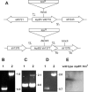
FIG. 1.
FIG. 1.
Insertional inactivation of the lepB1 and lepB2 genes in Synechocystis sp. strain PCC 6803. (A) Schematic maps of the chromosomal regions covering open reading frames sll0716 and slr1377. The restriction sites used for insertion of the kanamycin resistance cassette (Kmr) are indicated. The positions of the primers used for PCR analysis (forward primers F1, F2, and F3 and reverse primers R1, R2, and R3) are indicated by small arrows. (B to D) Segregation analyses of the lepB1::Kmr (B), lepB2::Kmr (C), and slr1378::Kmr (D) insertion mutants. Chromosomal DNA of wild-type Synechocystis sp. strain PCC 6803 (lanes 1) or the mutants (lanes 2) was amplified by PCR by using primers F1 and R1 (B), primers F2 and R2 (C), and primers F3 and R3 (D) and separated on 1% agarose gels. The sizes of relevant PCR products (in kilobases) are indicated on the right. (E) Western analysis of thylakoid proteins from wild-type and lepB1::Kmr mutant cells. Thylakoid proteins (100 μg of protein per lane) were separated by electrophoresis on SDS-10 to 17.5% polyacrylamide gradient gels, transferred to nylon membranes, and analyzed with antisera raised against LepB1 from Synechocystis sp. strain PCC 6803. For further details see the text.
FIG. 2.
FIG. 2.
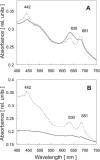
Light sensitivity of photosynthetic pigments in the lepB1::Kmr mutant. Cultures of wild-type Synechocystis sp. strain PCC 6803 (dotted line) and the lepB1::Kmr mutant (solid line) were grown under photomixotrophic conditions in dim light (5 microeinsteins m−2 s−1). The absorption spectra from identical amounts of cells were determined either directly (A) or after exposure of the cultures to a light intensity of 40 microeinsteins m−2 s−1 for 2 days (B). The absorption maxima for chlorophyll a (442 and 681 nm) and phycocyanin (630 nm) are indicated. rel. units, relative units.
FIG. 3.
FIG. 3.
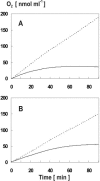
High light intensities lead to inactivation of photosynthetic oxygen evolution in the lepB1::Kmr mutant. The photosynthetic oxygen evolution by wild-type (dotted line) and lepB1::Kmr cultures (solid line) which were exposed to continuous light at an intensity of either 500 microeinsteins m−2 s−1 (A) or 100 microeinsteins m−2 s−1 (B) was determined for identical amounts of cells by using a Clark electrode.
FIG. 4.
FIG. 4.
Photosynthetic oxygen evolution by lepB1::Kmr mutant cells is inhibited by light stress. Cultures of wild-type Synechocystis sp. strain PCC 6803 (□) and the lepB1::Kmr mutant (○) propagated under photomixotrophic conditions in dim light (5 microeinsteins m−2 s−1) were exposed to light stress (1,000 microeinsteins m−2 s−1). Samples containing identical amounts of cells were taken at different times, and photosynthetic oxygen evolution was immediately determined for 5 min by using a Clark electrode. The value for each time is the average of at least five independent measurements. The error bars indicate the standard errors.
FIG. 5.
FIG. 5.
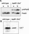
Western analysis of thylakoid proteins in wild-type and lepB1::Kmr cells. Thylakoid proteins isolated from cultures propagated under photomixotrophic conditions in dim light were separated by electrophoresis on SDS—10 to 17.5% polyacrylamide gradient gels, transferred to nylon membranes, and analyzed with antisera raised against PsbO (A) or cytochrome f (B) from spinach. The positions of the presumed precursor and mature proteins are indicated on the right. Lanes a, 50 μg of protein; lanes b, 100 μg of protein.
FIG. 6.
FIG. 6.
Two-dimensional analysis of total proteins from Synechocystis sp. strain PCC 6803. Total proteins were isolated from wild-type and lepB1::Kmr cells grown under photomixotrophic conditions in dim light and were separated in two dimensions by isoelectric focusing (pI 4.0 to 7.0) and SDS-polyacrylamide gel electrophoresis (10 to 17.5% polyacrylamide gradient gel). In each case, 200 μg of protein was loaded. Examples of gels obtained with proteins isolated from either wild-type cells (left panel) or lepB1::Kmr cells (right panel) are shown after staining with Coomassie brilliant blue. The numbers indicate protein spots that in five independent experiments always showed significant differences. These spots were recovered and analyzed by matrix-assisted laser desorption ionization—time of flight mass spectrometry (Table 2).

Similar articles

Cited by

References

    1. Anderson, S. L., and L. McIntosh. 1991. Light-activated heterotrophic growth of the cyanobacterium Synechocystis sp. strain PCC 6803: a blue-light-requiring process. J. Bacteriol. 173:2761-2767. - PMC - PubMed
    1. Barbrook, A. C., J. C. L. Packer, and C. J. Howe. 1993. Components of the protein translocation machinery in the thermophilic cyanobacterium Phormidium laminosum. Biochem. Biophys. Res. Commun. 197:874-877. - PubMed
    1. Berghöfer, J., and R. B. Klösgen. 1999. Two distinct translocation intermediates can be distinguished during protein transport by the TAT (delta pH) pathway across the thylakoid membrane. FEBS Lett. 460:328-332. - PubMed
    1. Bonnemain, C., C. Raynaud, H. Reglier-Poupet, I. Dubail, C. Frehel, M. A. Lety, P. Berche, and A. Charbit. 2004. Differential roles of multiple signal peptidases in the virulence of Listeria monocytogenes. Mol. Microbiol. 51:1251-1266. - PubMed
    1. Burnap, R. L., and L. A. Sherman. 1991. Deletion mutagenesis in Synechocystis sp. PCC6803 indicates that the Mn-stabilizing protein of photosystem II is not essential for O2 evolution. Biochemistry 30:440-446. - PubMed

Publication types

MeSH terms

LinkOut - more resources