. 2005 May;187(9):3133-8.
doi: 10.1128/JB.187.9.3133-3138.2005.

Plasmid-encoded regulator of extracellular proteases in Bacillus anthracis

Affiliations

Plasmid-encoded regulator of extracellular proteases in Bacillus anthracis

Arthur I Aronson et al. J Bacteriol. 2005 May.

Abstract

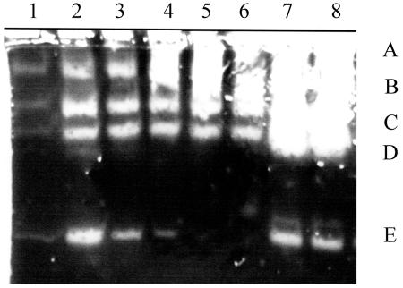
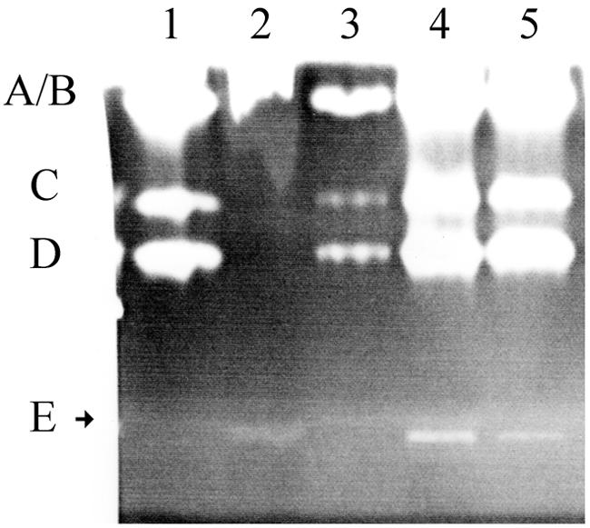
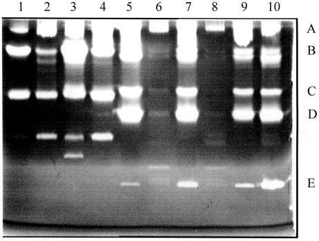
Bacillus anthracis Sterne cured of the pXO1 plasmid had enhanced secreted protease activity during the postexponential phase but no change in hemolytic or lecithinase activities. A zymogen profile revealed at least six proteases, including serine, metal, and perhaps cysteine types. There were similar amounts of protease secreted by the closely related species Bacillus cereus and Bacillus thuringiensis, but the patterns differed. Among the pXO1 plasmid-encoded proteins, there is a tetratricopeptide protein designated Cot43 that is related to the Rap proteins of Bacillus subtilis and the PlcR pleiotropic regulator of secreted enzymes and toxins in B. thuringiensis. A disruption of the cot43 gene resulted in overproduction of several proteases to a somewhat greater extent than in the plasmid-cured strain. Transformation of either of these strains with a clone of the cot43 gene resulted in the inhibition of accumulation of some of the proteases and induction of at least one. On the basis of lacZ fusions, transcription of the cot43 gene increased in late exponential cells at the time of protease accumulation. The expression of lacZ fusions to the upstream regions of two B. anthracis extracellular protease genes was greater in the strain with the disruption of cot43 than in the Sterne strain, indicating regulation at the level of transcription. In B. anthracis, a pXO1 plasmid-encoded protein directly modulates or indirectly regulates the transcription of genes for several chromosomally encoded extracellular proteases.


Figures

FIG. 1.
FIG. 1.
Zymogen profiles of a gel containing low-fat milk. Lanes 1 to 5 are supernatants from the Sterne strain grown in NSM at 37°C with samples removed at 60-min intervals, commencing at the end of growth. Phase-white endospores were present in >80% of the cells by sample 4. Lanes 6 to 8 are for the plasmid-free strain, St-13, sampled at the same time as lanes 2, 4, and 5. Bands labeled A to E are discussed in the text (Fig. 2). In all cases, 5 μl of the supernatants was loaded from cultures grown to a Klett value of 230 or the equivalent (see Materials and Methods).
FIG. 2.
FIG. 2.
Effect of inhibitors on extracellular proteases. Lane 1, 5 μl of the supernatant from the Sterne strain grown in NSM at 37°C and harvested as described for lane 4, Fig. 1; lane 2, addition of EGTA to a concentration of 2.5 mM prior to loading; lane 3, 2.5 mM PMSF; lane 4, 2.5 mM O-phenanthroline; lane 5, 2.5 mM E64. A molecular weight marker was a metalloprotease of 36 kDa from B. polymyxa (Sigma P-6141) which migrated a little bit more slowly than band E (Fig. 5).
FIG. 3.
FIG. 3.
Zymogen profiles with variations in strains, media, and temperature of growth. Lane 1, B. cereus T grown in G-tris medium at 30°C; lane 2, B. cereus T grown in NSM at 30°C; lane 3, B. cereus T grown at 37°C in G-tris; lane 4, B. cereus T grown at 37°C in NSM; lane 5, Sterne strain grown at 30°C in NSM; lane 6, Sterne strain grown at 30°C in BHI plus 0.5 M sorbitol; lane 7, Sterne strain grown at 37°C in NSM; lane 8, Sterne strain grown at 37°C in BHI plus 0.5 M sorbitol; lanes 9 and 10, Sterne strain grown at 37°C in NSM and samples incubated at 27°C for 30 min prior to loading (lane 9 supernatant from cells sampled 90 min earlier than the samples in the other lanes). Supernatants in lanes 1 to 8 and 10 are from samples taken 3 h after the end of growth (20 to 30% phase-dull spores in each). See legend to Fig. 1 for the amounts loaded.
FIG. 4.
FIG. 4.
Time course of the increase in the specific activity of β-galactosidase in the Sterne strain containing a lacZ fusion to the cot43 upstream region. Sampling and analysis were performed as described in Materials and Methods.
FIG. 5.
FIG. 5.
Enhanced protease in strains lacking Cot43. Lane 1, B. polymyxa metalloprotease (the lower band is 36 kDa); lane 2, Sterne strain with a disruption of the cot43 gene (strain d2); lane 3, strain UT44; lane 4, strain St-13; lane 5, Sterne strain; Lanes 6 to 9, the same strains used in lanes 2 to 5, respectively, but with cells harvested 90 min earlier. Strains were grown at 37°C in NSM until 90 min after the end of growth (lanes 6 to 9) or 180 min after the end of growth (lanes 2 to 5). Bands labeled A to E are as discussed in the legend to Fig. 2 and the text. Amounts loaded are as described in the legend to Fig. 1.
FIG. 6.
FIG. 6.
A clone of the cot43 gene regulates the intensity of the protease bands. Supernatants of strain St-13 (lanes 1, 4, and 7), strain St-13/cot43 (lanes 2, 5, and 8), and strain St-13/cot43P (lanes 3, 6, and 9) were used. Samples were removed during late exponential growth (lanes 1 to 3), at the end of growth (lanes 4 to 6), and 90 min later (lanes 7 to 9). Amounts were loaded as described in the legend to Fig. 1.
FIG. 7.
FIG. 7.
Differential effects of the presence of Cot43 on the zymogen profiles of a mutant with a disruption of the atxA gene. Lane 1, strain UM44; lane 2, strain UT53; lane 3, strain UT53/cot43P. All strains were grown at 37°C in NSM until 1 h after the end of exponential growth. Amounts were loaded as described in the legend of Fig 1.
FIG. 8.
FIG. 8.
Specific activities of β-galactosidase of a fusion of the upstream region of the immune inhibitor protease to lacZ in the Sterne strain and in mutant d2. Time zero is the end of growth, with phase-dull endospores present in >70% of the cells by 4 h.

References

    1. Agaisse, H., and D. Lereclus. 1994. Structural and functional analysis of the promoter region involved in full expression of the cryIIIA toxin gene of Bacillus thuringiensis. Mol. Microbiol. 13:97-107. - PubMed
    1. Agaisse H., M. Gominet, O. A. Okstad, A.-B. Kolsto, and D. Lereclus. 1999. PlcR is a pleiotropic regulator of extracellular virulence factor gene expression in Bacillus thuringiensis. Mol. Microbiol. 32:1043-1053. - PubMed
    1. Arantes, O., and D. Lereclus. 1991. Construction of cloning vectors for Bacillus thuringiensis. Gene 108:115-119. - PubMed
    1. Aronson, A. I., N. Angelo, and S. C. Holt. 1971. Regulation of extracellular protease production in Bacillus cereus T: characterization of mutants producing altered amounts of protease. J. Bacteriol. 106:1016-1025. - PMC - PubMed
    1. Bendtsen, J. D., H. Nielsen, G. von Heinje, and S. Brunek. 2004. Improved prediction of signal peptides: signal IP30. J. Mol. Biol. 340:783-795. - PubMed

Publication types

LinkOut - more resources