. 2005 May;187(9):3139-50.
doi: 10.1128/JB.187.9.3139-3150.2005.

Staphylococcus intermedius produces a functional agr autoinducing peptide containing a cyclic lactone

Affiliations

Staphylococcus intermedius produces a functional agr autoinducing peptide containing a cyclic lactone

Guangyong Ji et al. J Bacteriol. 2005 May.

Abstract

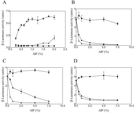
The agr system is a global regulator of accessory functions in staphylococci, including genes encoding exoproteins involved in virulence. The agr locus contains a two-component signal transduction module that is activated by an autoinducing peptide (AIP) encoded within the agr locus and is conserved throughout the genus. The AIP has an unusual partially cyclic structure that is essential for function and that, in all but one case, involves an internal thiolactone bond between a conserved cysteine and the C-terminal carboxyl group. The exceptional case is a strain of Staphylococcus intermedius that has a serine in place of the conserved cysteine. We demonstrate here that the S. intermedius AIP is processed by the S. intermedius AgrB protein to generate a cyclic lactone, that it is an autoinducer as well as a cross-inhibitor, and that all of five other S. intermedius strains examined also produce serine-containing AIPs.


Figures

FIG. 1.
FIG. 1.
Comparison of the AgrD amino acid sequences from S. aureus and S. intermedius. The amino acid sequences were analyzed by the Clone Manager software (Sci-Ed Software, Durham, NC). Dash lines represent gaps generated by the analysis program. The identical amino acid residues are dark shaded, and the AIP sequences in AgrDs are in boldface. The amino acid sequences of AgrD proteins can be accessed through the NCBI protein database under NCBI accession no. CAA36782 (S. aureus group I, Sa I) (22), AAB63265 (S. aureus group II, Sa II) (8), AAB63268 (S. aureus group III, Sa III) (8), AAG03056 (S. aureus group IV, Sa IV) (7), AY871105 (RN9161), AY871106 (RN9167), AY871107 (RN9169), AAL65836 (CCM5739) (2), AF346723 (RN9515), and AAS66746 (ATCC 29663). The amino acid sequences of AgrD proteins can be accessed through the NCBI protein database under NCBI accession no. CAA36782 (S. aureus group I, Sa I) (22), AAB63265 (S. aureus group II, Sa II) (8), AAB63268 (S. aureus group III, Sa III) (8), AAG03056 (S. aureus group IV, Sa IV) (7), AAC38294 (S. epidermidis, Se) (35), AAA71977 (S. lugdunensis, Sl) (34), and AAS66746 (S. intermedius ATCC 29663, Si).
FIG. 2.
FIG. 2.
agr self-activation by S. intermedius CCM5739 supernatant. (A) Northern blot analysis. The lyophilized residue from an equal volume of postexponential S. intermedius supernatant (plus S. intermedius) or broth (+BH) was added to an early-exponential-phase culture of CCM5739, and samples were drawn every 30 min for Northern blot hybridization analysis with an RNAIII-specific probe. (B) Growth curves. Cell density monitored turbidimetrically during the 4-h experiment is plotted versus time.
FIG. 3.
FIG. 3.
(A) agr inhibition by S. intermedius supernatants. To 50-μl aliquots of early-exponential-phase cultures (at cell densities of ∼100 Klett) of the four agr group-specific tester strains were added 10 μl of culture supernatants from the respective agr wild-type strains plus 0, 10, or 30 μl of a culture supernatant of the S. intermedius strain to be tested for inhibitory activity. Total volumes were made up to 100 μl with CYGP broth, and the plates were incubated for 90 min with shaking at 37°C. Then, 50 μl was transferred to a new microtiter plate, 50 μl of saturated nitrocefin was added, and the β-lactamase reaction was monitored kinetically, with the slope of the reaction over the first 5 min used to represent the enzyme activity. The rates in the presence of the S. intermedius supernatants were then normalized to the rate in the presence of activator alone. Hatched bars, 10 μl of S. intermedius supernatant; black bars, 30 μl of S. intermedius supernatant; shaded bars, control, activator only. (B) S. intermedius agr inhibition by S. aureus group I, II, and III AIPs. Culture supernatants were prepared from S. aureus strains (group I, RN6390B; group II, SA502A; and group III, RN8463). AIP activity assays with S. intermedius harboring pWP1004 as reporter cells were performed as described in Materials and Methods.
FIG. 4.
FIG. 4.
Interaction between AgrB and AgrD. Conditioned media were prepared from S. aureus GJ2035 expressing various combinations of AgrB and AgrD, and the AIP activities were measured by using S. intermedius containing pWP1004 as reporter cells. (A and B) Test for activation of S. intermedius agr by AIPs prepared from cells coexpressing S. aureus AgrB-I and AgrB-II or S. intermedius AgrB-Si and the S. intermedius AgrD-Si (A) and test for inhibition of S. intermedius agr by AIPs prepared from bacteria coexpressing the S. intermedius AgrB-Si and S. aureus AgrD-I, AgrD-II, or AgrD-III (B). Reporter cells grown in the absence of AIP were used as controls. Values are means from three independent experiments with standard errors as indicated.
FIG. 5.
FIG. 5.
Inability of AgrB-I to process a serine-containing AgrD-I mutant. Using a cloned agrD-I derivative, the cysteine codon, TGT, was replaced by a serine codon, AGT, and the mutant agrD was cloned into a vector between N- and C-terminal His6 tags. The resulting construct was tested in vivo in the presence or absence of an agrB-I- containing plasmid and compared to the native agrD-I, also containing the N- and C-terminal His tags, for the production of agr-activating or -inhibiting substances by using agr reporter strains with a β-lactamase readout. (A) For activation tests, the culture was grown for 90 min in the presence of the supernatant to be tested for activation and then assayed. (B) For inhibition tests, a sample of a cognate supernatant (activator) (i.e., group II supernatant for the group II reporter and group III supernatant for the group III reporter) was added at the same time as the supernatant to be tested for inhibition, and the culture then grown for 90 min and assayed for β-lactamase. For each of the five sets of tests shown, the columns are labeled as follows: A, wild-type AgrD; B, AgrD, C28S; C, wild-type AgrD plus AgrB-I; D, AgrD, C28S plus AgrB-I [the reporters were RN6390B(pRN6683) for group I, SA502A(pRN6683) for group II, and RN8463(pRN6683) for group III as described previously (8, 9)].
FIG. 6.
FIG. 6.
Processing of the wild-type and the serine-to-cysteine mutant AgrD-Si by AgrB-Si. (A to D) AIP activity assays. S. aureus cells were grown and induced. After centrifugation, the culture supernatants were used as the conditioned media (either concentrated or diluted with CYGP medium) to perform either AIP activation assays with S. intermedius containing pWP1004 as reporter cells (A) or AIP inhibition assays with RN6390B(pRN6683) (S. aureus group I) (B), SA502A(pRN6683) (S. aureus group II) (C), and RN8463(pRN6683) (S. aureus group III) (D) reporter cells. Undiluted conditioned media were equal to 100% AIP. Conditioned media were prepared from cultures of cells coexpressing AgrB-Si and the wild-type AgrD-Si (•), AgrD-Si(S27C) (▴), or AgrD-Si(S27A) (▪). Values are means from three independent experiments with the standard errors as indicated. (E) Western blot hybridization analysis. S. aureus cells coexpressing AgrB-Si and wild-type AgrD-Si, AgrD-Si(S27C), or AgrD-Si(S27A) were grown and induced, and the cell cultures were mixed with ethanol-acetone (1:1). The mixtures were centrifuged, and the cells were washed and lysed. Whole-cell lysates were separated by SDS-polyacrylamide gel electrophoresis and transferred to nitrocellulose membranes. The membranes were then probed with an anti-T7 tag monoclonal antibody.
FIG. 6.
FIG. 6.
Processing of the wild-type and the serine-to-cysteine mutant AgrD-Si by AgrB-Si. (A to D) AIP activity assays. S. aureus cells were grown and induced. After centrifugation, the culture supernatants were used as the conditioned media (either concentrated or diluted with CYGP medium) to perform either AIP activation assays with S. intermedius containing pWP1004 as reporter cells (A) or AIP inhibition assays with RN6390B(pRN6683) (S. aureus group I) (B), SA502A(pRN6683) (S. aureus group II) (C), and RN8463(pRN6683) (S. aureus group III) (D) reporter cells. Undiluted conditioned media were equal to 100% AIP. Conditioned media were prepared from cultures of cells coexpressing AgrB-Si and the wild-type AgrD-Si (•), AgrD-Si(S27C) (▴), or AgrD-Si(S27A) (▪). Values are means from three independent experiments with the standard errors as indicated. (E) Western blot hybridization analysis. S. aureus cells coexpressing AgrB-Si and wild-type AgrD-Si, AgrD-Si(S27C), or AgrD-Si(S27A) were grown and induced, and the cell cultures were mixed with ethanol-acetone (1:1). The mixtures were centrifuged, and the cells were washed and lysed. Whole-cell lysates were separated by SDS-polyacrylamide gel electrophoresis and transferred to nitrocellulose membranes. The membranes were then probed with an anti-T7 tag monoclonal antibody.
FIG. 7.
FIG. 7.
Hemolytic patterns. Cultures to be tested were grown overnight on GL agar, cross-streaked on sheep blood agar against a culture of RN4220, incubated overnight at 37°C, and then incubated for 6 h at 4°C. The patterns can be interpreted as follows: the “hot-cold” beta-hemolysin appears as a partially turbid zone, as seen with RN4220, which produces only beta-hemolysin. Delta-hemolysin is synergistic with beta-hemolysin and is seen as a clearing where the two hemolysins intersect; this is best seen with RN9515 (top left). Coproduction of beta-hemolysin and delta-hemolysin is seen as a clearing next to the streak within a wider beta-hemolysin zone (best illustrated with RN9169, bottom left) and, less strongly, with RN9167 (bottom right). RN9161 produces only delta-hemolysin and quite weakly. RN6734 produces a very strong alpha-hemolysin zone, as shown by the characteristic antagonism between alpha-hemolysin and beta-hemolysin. It also produces delta-hemolysin, as shown by the clearer zone where the beta-hemolysin and delta-hemolysin zones intersect. RN7206 produces a very weak alpha-hemolysin zone, as shown by its inhibition by beta-hemolysin. The CCM5739 (labeled RN9423) pattern could represent a beta-hemolysin zone equivalent to that of RN9169 plus a very strong delta-hemolysin zone, which obscures that of beta-hemolysin. The interaction of CCM5739 (labeled RN9423) with the RN4220 beta-hemolysin zone is very atypical and is not interpretable according to our understanding of the activities of the S. aureus hemolysins.

Similar articles

Cited by

References

    1. Becker, K., B. Keller, C. von Eiff, M. Bruck, G. Lubritz, J. Etienne, and G. Peters. 2001. Enterotoxigenic potential of Staphylococcus intermedius. Appl. Environ. Microbiol. 67:5551-5557. - PMC - PubMed
    1. Dufour, P., S. Jarraud, F. Vandenesch, T. Greenland, R. P. Novick, M. Bes, J. Etienne, and G. Lina. 2002. High genetic variability of the agr locus in Staphylococcus species. J. Bacteriol. 184:1180-1186. - PMC - PubMed
    1. Fitton, J. E., A. Dell, and W. V. Shaw. 1980. The amino acid sequence of the delta haemolysin of Staphylococcus aureus. FEBS Lett. 115:209-212. - PubMed
    1. Girard, C., and R. Higgins. 1999. Staphylococcus intermedius cellulitis and toxic shock in a dog. Can. Vet. J. 40:501-502. - PMC - PubMed
    1. Janzon, L., and S. Arvidson. 1990. The role of the delta-lysin gene (hld) in the regulation of virulence genes by the accessory gene regulator (agr) in Staphylococcus aureus. EMBO J. 9:1391-1399. - PMC - PubMed

Publication types

MeSH terms

Associated data

LinkOut - more resources