. 2005 May;187(9):3238-48.
doi: 10.1128/JB.187.9.3238-3248.2005.

Molecular determinants for PspA-mediated repression of the AAA transcriptional activator PspF

Affiliations

Molecular determinants for PspA-mediated repression of the AAA transcriptional activator PspF

Sarah Elderkin et al. J Bacteriol. 2005 May.

Abstract

The Escherichia coli phage shock protein system (pspABCDE operon and pspG gene) is induced by numerous stresses related to the membrane integrity state. Transcription of the psp genes requires the RNA polymerase containing the sigma(54) subunit and the AAA transcriptional activator PspF. PspF belongs to an atypical class of sigma(54) AAA activators in that it lacks an N-terminal regulatory domain and is instead negatively regulated by another regulatory protein, PspA. PspA therefore represses its own expression. The PspA protein is distributed between the cytoplasm and the inner membrane fraction. In addition to its transcriptional inhibitory role, PspA assists maintenance of the proton motive force and protein export. Several lines of in vitro evidence indicate that PspA-PspF interactions inhibit the ATPase activity of PspF, resulting in the inhibition of PspF-dependent gene expression. In this study, we characterize sequences within PspA and PspF crucial for the negative effect of PspA upon PspF. Using a protein fragmentation approach, we show that the integrity of the three putative N-terminal alpha-helical domains of PspA is crucial for the role of PspA as a negative regulator of PspF. A bacterial two-hybrid system allowed us to provide clear evidence for an interaction in E. coli between PspA and PspF in vivo, which strongly suggests that PspA-directed inhibition of PspF occurs via an inhibitory complex. Finally, we identify a single PspF residue that is a binding determinant for PspA.


Figures

FIG. 1.
FIG. 1.
PspA fragments. (A) Schematic showing (to scale) the predicted four helical domains (HD1 to -4) of E. coli PspA using the COILS program. (B) Coomassie blue-stained 18% SDS-PAGE gel showing the PspA fragments purified as six-His fusion proteins.
FIG. 2.
FIG. 2.
Interaction between PspA fragments and the AAA domain of PspF (PspF1-275). Coomassie-stained native gel showing complex formation between 5 μM PspF1-275 and increasing amounts of PspA or PspA fragments (5, 10, and 20 μM). Migration properties of PspA and PspA fragments (20 μM) in the absence (−) of PspF are shown in lanes 2, 6, 10, 14, 18, and 22.
FIG. 3.
FIG. 3.
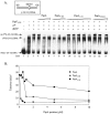
Effects of PspA fragments on PspF1-275 functioning. (A) Native gel showing the effects of PspA fragments on nucleotide-dependent isomerization of σ54 bound to (−11/−12) DNA probe by PspF1-275 when PspA fragments were preincubated with PspF1-275. Reaction mixtures contained (+) 16 nM of 32P-end-labeled S. meliloti nifH promoter (−11/−12) DNA probe, 1 μM of σ54, 4 mM dGTP, 1 μM of PspF1-275, and increasing concentrations of PspA or PspA fragments (1, 2, and 3 μM for PspA, Psp1-186, and PspA110-222; 1, 2, 3, 5, and 10 μM for PspA1-110). The isomerized complex is marked ss σ54/(−11/−12) DNA. Complexes were detected by PhosphorImager analysis. (B) Effects of PspA fragments on PspF ATPase activity. The ATPase activity of PspF was measured by incubating 500 nM PspF with [α-32P]ATP for 30 min, followed by separation of [α-32P]ATP and [α-32P]ADP on thin-layer chromatography and was quantified by PhosphorImager analysis. The graph shows the amount of ATP turnover of PspF when preincubated with increasing concentrations of PspA or PspA fragments (PspA1-186 and PspA1-110s) 10 min before the addition of [α-32P]ATP to start the ATP hydrolysis reaction. (C) Native gel showing the effects of the combination of PspA fragments PspA110-222 and PspA1-110 on nucleotide-dependent isomerization of σ54 bound to (−11/−12) DNA probe by PspF1-275. σ isomerization reactions were conducted as for panel A. When present (+), PspA was at 3 μM (lane 4). The reactions in lanes 5 to 9 were done using 3 μM PspA1-110. In lanes 6 to 8, increasing concentrations of PspA110-222 (1, 2, and 3 μM) were added to 3 μM of PspA1-110. Lane 9 and lane 15 represent the addition of PspA fragment storage buffer and BSA, respectively, substituted for 3 μM PspA110-222. The reactions in lanes 10 to 14 were done using 3 μM PspA110-222. In lanes 11 to 13, increasing concentrations of PspA1-110 (1, 2, and 3 μM) were added to 3 μM of PspA110-222. Lane 14 represents the addition of storage buffer substituted for 3 μM PspA1-110.
FIG. 3.
FIG. 3.
Effects of PspA fragments on PspF1-275 functioning. (A) Native gel showing the effects of PspA fragments on nucleotide-dependent isomerization of σ54 bound to (−11/−12) DNA probe by PspF1-275 when PspA fragments were preincubated with PspF1-275. Reaction mixtures contained (+) 16 nM of 32P-end-labeled S. meliloti nifH promoter (−11/−12) DNA probe, 1 μM of σ54, 4 mM dGTP, 1 μM of PspF1-275, and increasing concentrations of PspA or PspA fragments (1, 2, and 3 μM for PspA, Psp1-186, and PspA110-222; 1, 2, 3, 5, and 10 μM for PspA1-110). The isomerized complex is marked ss σ54/(−11/−12) DNA. Complexes were detected by PhosphorImager analysis. (B) Effects of PspA fragments on PspF ATPase activity. The ATPase activity of PspF was measured by incubating 500 nM PspF with [α-32P]ATP for 30 min, followed by separation of [α-32P]ATP and [α-32P]ADP on thin-layer chromatography and was quantified by PhosphorImager analysis. The graph shows the amount of ATP turnover of PspF when preincubated with increasing concentrations of PspA or PspA fragments (PspA1-186 and PspA1-110s) 10 min before the addition of [α-32P]ATP to start the ATP hydrolysis reaction. (C) Native gel showing the effects of the combination of PspA fragments PspA110-222 and PspA1-110 on nucleotide-dependent isomerization of σ54 bound to (−11/−12) DNA probe by PspF1-275. σ isomerization reactions were conducted as for panel A. When present (+), PspA was at 3 μM (lane 4). The reactions in lanes 5 to 9 were done using 3 μM PspA1-110. In lanes 6 to 8, increasing concentrations of PspA110-222 (1, 2, and 3 μM) were added to 3 μM of PspA1-110. Lane 9 and lane 15 represent the addition of PspA fragment storage buffer and BSA, respectively, substituted for 3 μM PspA110-222. The reactions in lanes 10 to 14 were done using 3 μM PspA110-222. In lanes 11 to 13, increasing concentrations of PspA1-110 (1, 2, and 3 μM) were added to 3 μM of PspA110-222. Lane 14 represents the addition of storage buffer substituted for 3 μM PspA1-110.
FIG. 4.
FIG. 4.
Western blot analysis of LexAwtDBD-PspA fragment fusion proteins. LexAwtDBD-PspA fragment fusions were expressed in E. coli SU202 using 1 mM IPTG. An immunoblot of crude lysates of IPTG-induced cultures (OD600, ∼0.5) probed with anti-LexA antiserum (Invitrogen) is shown. The calculated molecular masses (in kilodaltons) for the LexA fusions are in parentheses: lane 1, LexA-PspA (34.6); lane 2, LexA-PspA1-186 (30.4); lane 3, LexA-PspA1-110 (21.5); lane 4, LexA-PspA1-67 (16.5); lane 5, LexA-PspA67-222 (27); lane 6, LexA-PspA110-222 (22); lane 7, LexA-PspA67-186 (22.9).
FIG. 5.
FIG. 5.
Theoretical atomic model of a PspF1-275 hexamer highlighting the probable location of W56. The model was prepared by submitting the sequence of the PspF AAA domain to the 3D-JIGSAW comparative-modeling server (http://www.bmm.icnet.uk/servers/3djigsaw/). The server builds three-dimensional models for proteins based on homologues of known structures (2). Theoretical coordinates for residues P31 to V237 were obtained based on the crystal structure of NtrC1 (34) (Protein Data Base [PDB] entry, 1NY5) with 95% accuracy. The high accuracy of the model is linked to the high sequence identity between PspF and NtrC1 sequences for the region of interest: 50.5%. Because the AAA domain of p97 crystallized as a hexamer, the model was superimposed onto the D1 structure of p97, including residues 200 to 458 (52) (PDB entry, 1E32), using the computer program CCP4-GUI. An accurate superposition was obtained due to the high secondary-structure conservation among AAA domains. A theoretical PspF hexamer was then generated using the CCP4-GUI. The figures were prepared using Pymol (http://pymol.sourceforge.net/) and POV-Ray (http://www.povray.org/). (A) Top view of a theoretical PspF1-275 hexamer shown in a grey cartoon representation. W56 in individual monomers is shown in a red-sphere representation. The probable locations of two of the six W56s are further highlighted using red circles. (B) Side view of the same hexamer with the putative location of one W56 highlighted using a red circle.
FIG. 6.
FIG. 6.
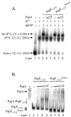
PspF1-275W56A escapes the inhibitory actions of PspA in vitro. (A) Native gel showing the effects of PspA on nucleotide-dependent isomerization of σ54 bound to (−11/−12) DNA probe by either PspF1-275 or PspF1-275W56A (1 μM) when increasing PspA fragments (2 and 3 μM) were preincubated with PspF1-275 proteins. Reactions were as in Fig. 3, and complexes were detected by PhosphorImager analysis. (B) Coomassie-stained native gel showing complex formation between PspA and either wild-type PspF1-275 or mutant PspF1-275W56A. Binding reactions were conducted with 5 μM PspF1-275 (lanes 2 to 5) or PspF1-275W56A (lanes 6 to 9) with increasing concentrations of PspA (0, 5, 10, and 15 μM).
FIG. 7.
FIG. 7.
In vivo effect of PspA on PspF1-275W56A-dependent activation of the pspA promoter. E. coli strain MVA4 carrying a chromosomal fusion, pspA::lacZ, was transformed with compatible plasmids pPB9, carrying pspA under control of placUV5, and either pPB8-WT (A) or pPB8-W56A (B), carrying wild-type pspF and mutant pspFW56A, respectively, under control of pBAD. Overnight cultures were diluted 100-fold, and cultures were grown at 37°C in LB medium supplemented with 0.2% glucose (to minimize the leaky expression from the placUV5 and pBAD promoters). After 3 h, overexpression of PspA and PspF proteins were induced by adding 1 mM IPTG and 0.2% arabinose for 1 h, and β-galactosidase activities were determined as described in Materials and Methods. The error bars indicate standard deviations.

Similar articles

Cited by

References

    1. Aseeva, E., F. Ossenbuhl, L. A. Eichacker, G. Wanner, J. Soll, and U. C. Vothknecht. 2004. Complex formation of Vipp1 depends on its alpha-helical PspA-like domain. J. Biol. Chem. 279:35535-35541. - PubMed
    1. Bates, P. A., L. A. Kelley, R. M. MacCallum, and M. J. E. Sternberg. 2001. Enhancement of protein modelling by human intervention in applying the automatic programs 3D-JIGSAW and 3D-PSSM. Proteins 5(Suppl.):39-46. - PubMed
    1. Beloin, C., J. Valle, P. Latour-Lambert, P. Faure, M. Kzreminski, D. Balestrino, J. A. Haagensen, S. Molin, G. Prensier, B. Arbeille, and J.-M. Ghigo. 2004. Global impact of mature biofilm lifestyle on Escherichia coli K-12 gene expression. Mol. Microbiol. 51:659-674. - PubMed
    1. Bogan, A. A., and K. S. Thorn. 1998. Anatomy of hot spots in protein interfaces. J. Mol. Biol. 280:1-9. - PubMed
    1. Bordes, P., S. R. Wigneshweraraj, J. Schumacher, X. Zhang, M. Chaney, and M. Buck. 2003. The ATP hydrolyzing transcription activator phage shock protein F of Escherichia coli: identifying a surface that binds σ54. Proc. Natl. Acad. Sci. USA 100:2278-2283. - PMC - PubMed

Publication types

MeSH terms

LinkOut - more resources