. 2005 Apr;2(4):e103.
doi: 10.1371/journal.pmed.0020103. Epub 2005 Apr 26.

Differentiation of insulin-producing cells from human neural progenitor cells

Affiliations

Differentiation of insulin-producing cells from human neural progenitor cells

Yuichi Hori et al. PLoS Med. 2005 Apr.

Abstract

Background: Success in islet-transplantation-based therapies for type 1 diabetes, coupled with a worldwide shortage of transplant-ready islets, has motivated efforts to develop renewable sources of islet-replacement tissue. Islets and neurons share features, including common developmental programs, and in some species brain neurons are the principal source of systemic insulin.

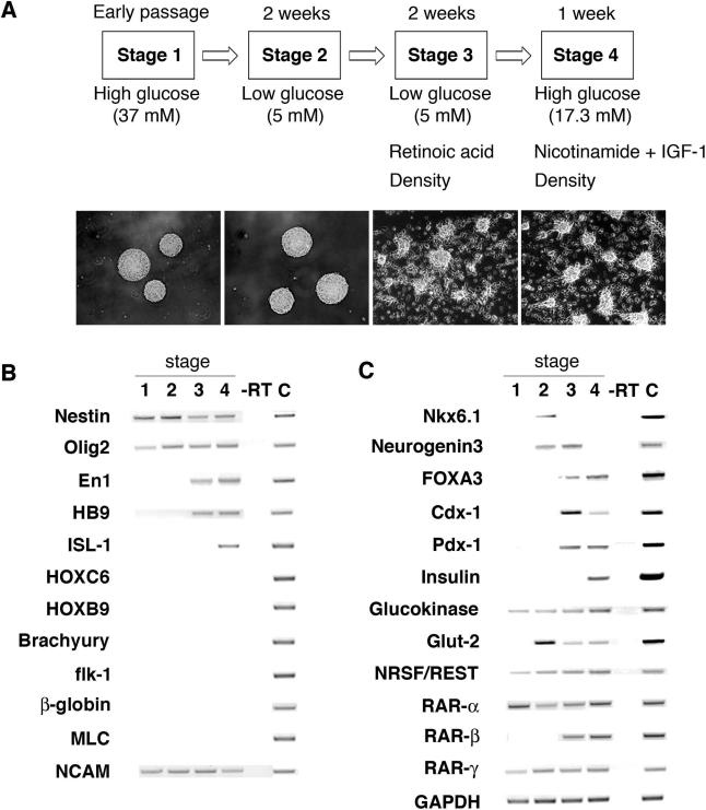
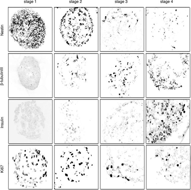
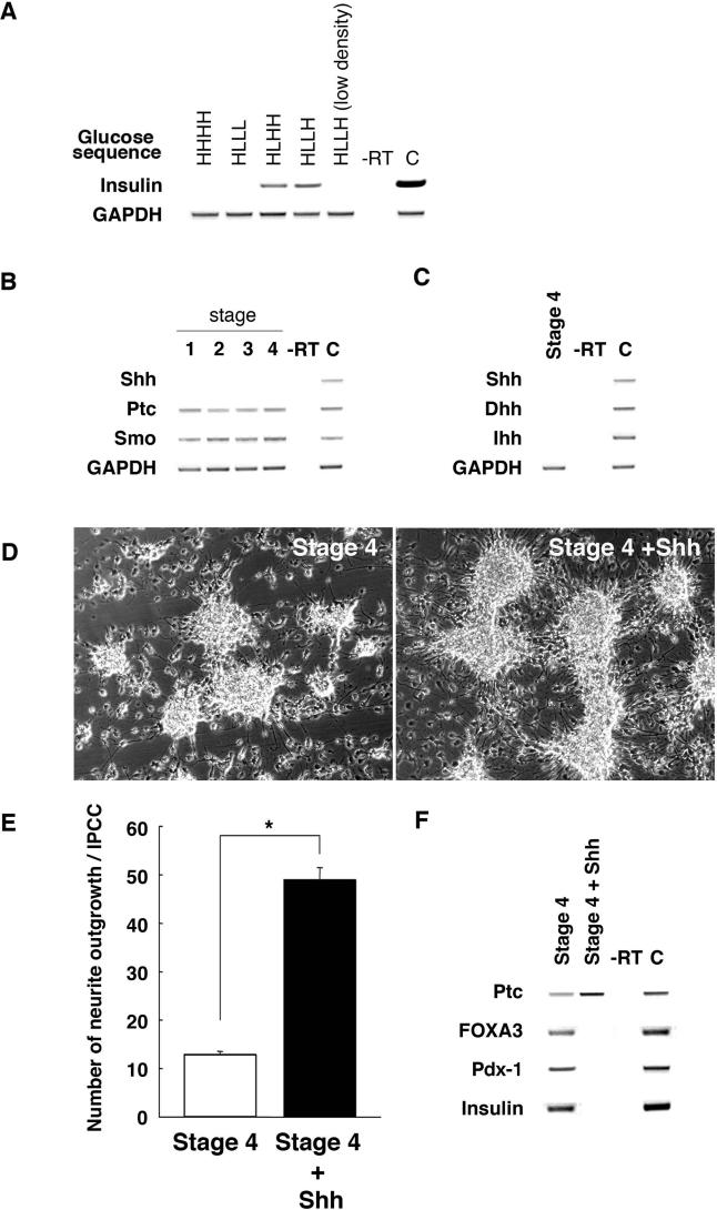
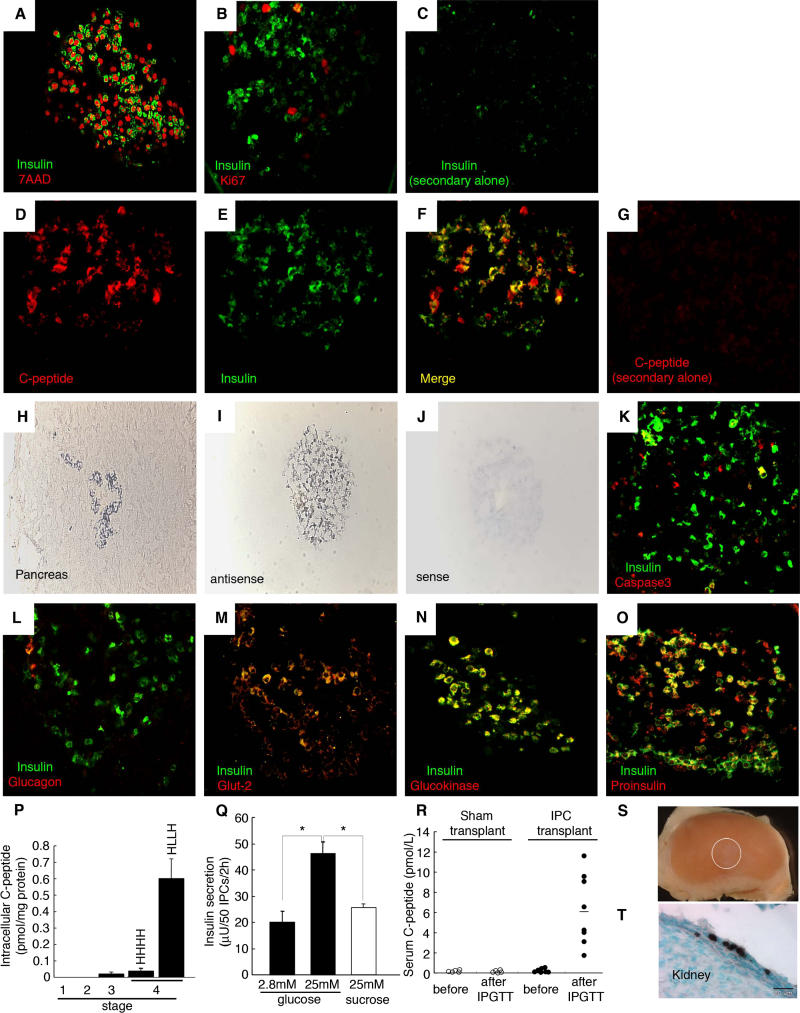
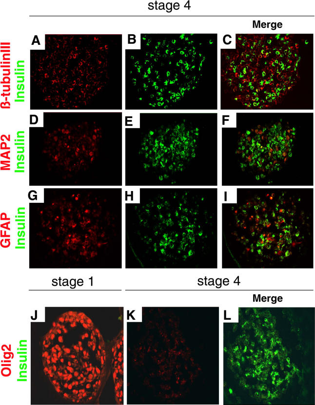
Methods and findings: Here we show that brain-derived human neural progenitor cells, exposed to a series of signals that regulate in vivo pancreatic islet development, form clusters of glucose-responsive insulin-producing cells (IPCs). During in vitro differentiation of neural progenitor cells with this novel method, genes encoding essential known in vivo regulators of pancreatic islet development were expressed. Following transplantation into immunocompromised mice, IPCs released insulin C-peptide upon glucose challenge, remained differentiated, and did not form detectable tumors.

Conclusion: Production of IPCs solely through extracellular factor modulation in the absence of genetic manipulations may promote strategies to derive transplantable islet-replacement tissues from human neural progenitor cells and other types of multipotent human stem cells.


Conflict of interest statement

Competing Interests: SK has a consulting relationship with and owns shares of the company StemCells.

Figures

Figure 1
Figure 1. Development of Cells Expressing Islet Markers from Human Neural Progenitor Cells
(A) Outline of four-stage differentiation protocol, essential factor manipulations at each stage, and stage-specific cell cluster morphology. Original magnification, 100×. (B) RT-PCR analysis of gene expression by stages 1–4 cell clusters, in stage 4 samples with RT omitted (–RT) and in human fetal spinal cord as a control (“C”). (C) RT-PCR analysis of expression during stages 1–4 of pancreatic and endodermal markers. Fetal pancreas RNA served as a control (“C”).
Figure 2
Figure 2. Immunohistochemical Detection of Neural Markers and Insulin during Stages 1–4 of Human Neural Progenitor Cell Differentiation
Immunofluorescent images were obtained by confocal microscopy and are representative of at least ten samples for each antibody. We detected insulin in only stage 4 IPCs, consistent with RT-PCR results. Distribution of insulin staining is localized in the cytoplasm. Immunofluorescent detection of Ki67, a nuclear marker of proliferating cells, showed stage 4 IPCs are predominantly non-proliferating, similar to mature pancreatic islets. Original magnification, 630×.
Figure 3
Figure 3. The Sequence of Glucose Concentration Changes, Cell Density, and Absence of Hh Signals Are Essential for Development of IPCs
(A) RT-PCR analysis of insulin gene expression for different glucose concentrations and cell densities during stages 2–4. High glucose is designated “H”; low glucose designated “L.” Thus, maintenance of glucose at high concentration in stages 1–4 is abbreviated as “HHHH”; exposure to high glucose in stage 1, followed by reduction of glucose in stages 2–4 is abbreviated “HLLL”; and so forth. Cell clusters analyzed here were cultured at 40–80 clusters/cm2 except where indicated (“low density”). Fetal pancreas RNA served as a control (“C”). (B) RT-PCR analysis of Hh signaling factors during stages 1–4 and in fetal spinal cord as a control (“C”). (C) RT-PCR analysis of stage 4 IPC expression of Shh, Desert hedgehog (Dhh), and Indian hedgehog (Ihh). Control (“C”) is a spinal cord sample. (D) Neurite outgrowth in control stage 4 IPC clusters, and in stage 4 IPC clusters exposed to 300 nM Shh. Original magnification, 100×. (E) Quantification of neurite outgrowth per control or Shh-treated stage 4 IPC cluster. *, P < 0.001. (F) RT-PCR analysis of IPC cluster expression of Ptc, FoxA3, Pdx1, and insulin following treatment with Shh at stage 4. Control (“C”) is fetal pancreas sample.
Figure 4
Figure 4. Stage 4 IPCs Express Characteristic Pancreatic β-Cell Markers and Are Glucose-Responsive
(A–C) Immunofluorescent images of stage 4 IPCs were obtained by confocal microscopy and are representative of at least ten samples for each probe. Shown is simultaneous immunofluorescent staining of insulin and either the nuclear marker 7AAD (A) or Ki67 (B). (C) Lack of immunostaining upon omission of the anti-insulin primary antibody. (D–G) Detection of insulin C-peptide in cells stained by anti-insulin antibodies. There is complete overlap between immunostained C-peptide and insulin in the cytoplasm. (H and I) Representative in situ hybridization with antisense riboprobes specific for human insulin on sectioned human fetal pancreatic islet cells (H) or a stage 4 IPC cluster (I). (J) Sense control probe in situ hybridization on a stage 4 IPC cluster. (K–O) Simultaneous immunofluorescent detection of insulin and cleaved caspase-3 (K), glucagon (L), Glut-2 (M), glucokinase (N), or proinsulin (O). (P) Intracellular C-peptide content of IPCs during stages 1–4 by human C-peptide-specific ELISA. (Q) In vitro secretion assay of stage 4 IPCs. Insulin release followed a step increase from 2.8 mM to 25 mM glucose, but not following exposure to 25 mM sucrose, which served as an osmotic control. *, p < 0.001. (R) Serum human C-peptide detection 2-wk after sham or IPC cluster transplantation in NOD scid mice. Samples were obtained before or 30 min after intraperitoneal glucose challenge (IPGTT). (S) Lack of tumor growth after renal transplantation of stage 4 IPCs (graft indicated by white circle). (T) Immunohistochemical detection of human C-peptide expression in stage 4 IPCs (cells stained brown) 2-wk after engraftment; bar is 20 μm. Original magnification of (H–J) and (T) was 250×. All other photomicrographs' original magnification was 630×.
Figure 5
Figure 5. Stage 4 IPCs Do Not Express Markers of Differentiated Neural Cells
(A–C) Immunohistochemical detection of β-tubulin III (A) and insulin (B) in stage 4 cells, and a merge of both images (C). (D–F) Immunohistochemical detection of MAP2 (D) and insulin (E) in stage 4 cells, and a merge of both images (F). (G–I) Immunohistochemical detection of GFAP (G) and insulin (H) in stage 4 cells, and a merge of both images (I). (J–L) Immunohistochemical detction of Olig2 at stage 1 (J) and stage 4 (K) and merged view of Olig2+ and insulin+ cells at stage 4 (L). Original magnification was 630×.

Similar articles

Cited by

References

    1. Shapiro AM, Lakey JR, Ryan EA, Korbutt GS, Toth E, et al. Islet transplantation in seven patients with type 1 diabetes mellitus using a glucocorticoid-free immunosuppressive regimen. N Engl J Med. 2000;242:230–238. - PubMed
    1. Edlund H. Pancreatic organogenesis—Developmental mechanisms and implications for therapy. Nat Rev Genet. 2002;3:524–532. - PubMed
    1. Rulifson EJ, Kim SK, Nusse R. Ablation of insulin-producing neurons in flies: Growth and diabetic phenotypes. Science. 2002;296:1118–1120. - PubMed
    1. Ikeya T, Galic M, Belawat P, Nairz K, Hafen E. Nutrient-dependent expression of insulin-like peptides from neuroendocrine cells in the CNS contributes to growth regulation in Drosophila . Curr Biol. 2002;12:1293–1300. - PubMed
    1. Deltour L, Leduque P, Blume N, Madsen O, Dubois P, et al. Differential expression of the two nonallelic proinsulin genes in the developing mouse embryo. Proc Natl Acad Sci U S A. 1993;90:527–533. - PMC - PubMed

Publication types

Associated data