. 2005 May 10;335(2):165-76.
doi: 10.1016/j.virol.2005.02.017.

Expression, purification, and characterization of SARS coronavirus RNA polymerase

Affiliations

Expression, purification, and characterization of SARS coronavirus RNA polymerase

Ao Cheng et al. Virology. .

Abstract

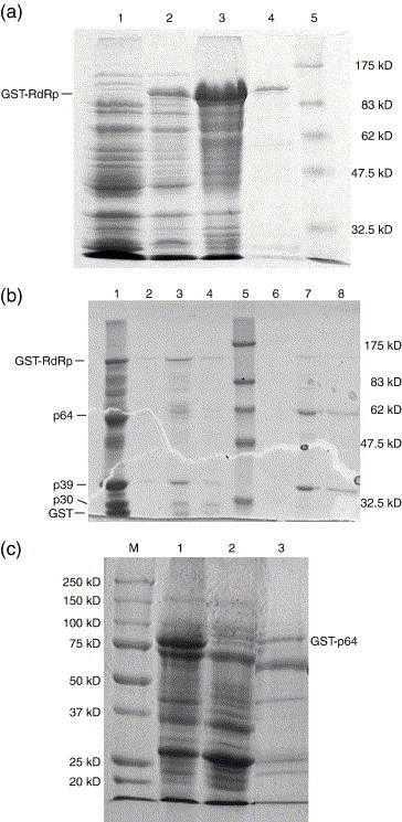
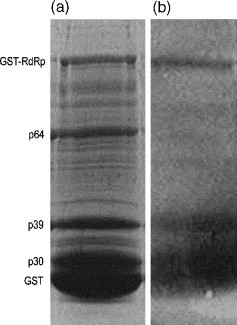
The RNA-dependent RNA polymerase (RdRp) of SARS coronavirus (SARS-CoV) is essential for viral replication and a potential target for anti-SARS drugs. We report here the cloning, expression, and purification of the N-terminal GST-fused SARS-CoV RdRp and its polymerase catalytic domain in Escherichia coli. During purification, the full-length GST-RdRp was found to cleave into three main fragments: an N-terminal p12 fragment, a middle p30 fragment, and a C-terminal p64 fragment comprising the catalytic domain, presumably due to bacterial proteases. Biochemical assays show that the full-length GST-RdRp has RdRp activity and the p64 and p12 fragments form a complex that exhibits comparable RdRp activity, whereas the GST-p64 protein has no activity, suggesting that the p12 domain is required for polymerase activity possibly via involvement in template-primer binding. Nonnucleoside HIV-1 RT inhibitors are shown to have no evident inhibitory effect on SARS-CoV RdRp activity. This work provides a basis for biochemical and structural studies of SARS-CoV RdRp and for development of anti-SARS drugs.


Figures

Fig. 1
Fig. 1
Expression of SARS-CoV RdRp. (a) Supernatant of the cell lysates. (b) Pellet of the cell lysates. Protein expression was carried out in the Origami (DE3) cells and induced at different IPTG concentrations and temperatures. Lanes 1–3: different IPTG concentrations of 0.1, 0.5, and 1 mM, respectively, at 37 °C for 3 h; lane 4: molecular mass standards; lanes 5–7: different IPTG concentrations of 0.1, 0.5, and 1 mM, respectively, at 28 °C for 3 h; and lanes 8–10: different IPTG concentrations of 0.1, 0.5, and 1 mM, respectively, at 24 °C for 3 h. The protein samples were analyzed in a 15% SDS-PAGE gel stained with Coomassie blue.
Fig. 2
Fig. 2
Purification of SARS-CoV RdRp. (a) Purification of GST-RdRp with the glutathione Sepharose 4B column. Lane 1: whole cell lysate without the expression vector; lane 2: supernatant; lane 3: pellet; lane 4: elution fraction; and lane 5: molecular mass standards. (b) Purification of GST-RdRp with the polyA Sepharose 4B column. Lane 1: the cleavage mixture of the protein sample after purification with the glutathione Sepharose 4B column in 5 days; lanes 2–4: flow-through fractions; lane 5: molecular mass standards; and lanes 6–8: elution fractions. (c) Purification of GST-p64 with the glutathione Sepharose 4B column. M: molecular mass standards; lane 1: pellet; lane 2: flow-through fraction; and lane 3: elution fraction.
Fig. 3
Fig. 3
Characterization of the p64/p12 complex. After purification with the glutathione Sepharose 4B column, the full-length GST-RdRp protein sample was stored at 4 °C for 5 days. One portion was treated with thrombin for the cleavage of GST and another was not treated with thrombin. Both samples were further purified with the polyA Sepharose 4B column. (a) SDS-PAGE of the purified protein samples without (left panel) and with thrombin treatment (right panel). (b) Native PAGE of GST (left panel) and the purified protein sample with thrombin treatment (right panel). (c) IEF gel of GST (left panel) and the purified protein sample with thrombin treatment (right panel).
Fig. 4
Fig. 4
Western blot analysis of the cleavage mixture of the full-length GST-RdRp. After purification with the glutathione Sepharose 4B column the protein sample was stored at 4 °C for 5 days. (a) SDS-PAGE of the protein sample. (b) Western blot of the protein sample. The gel shows that the full-length GST-RdRp and the p39 fragment contain a GST tag while the p64 fragment does not.
Fig. 5
Fig. 5
Schematic diagram showing the cleavage sites and compositions of the full-length SARS-CoV RdRp. SARS-CoV RdRp consists of 932 residues and comprises three domains. The N-terminal p12 domain consists of residues 1 to 110; the middle p30 domain comprises residues 111 to 368, and the C-terminal p64 domain contains residues 369 to 932. The cleavage site between the p12 and p30 domains is at M110–V111 and the cleavage site between the p30 and p64 domains is at F368–K369. p64 comprises the polymerase catalytic domain which contains three strictly conserved aspartates (Asp618, Asp760, and Asp761) that form the polymerase active site.

Similar articles

Cited by

References

    1. Anand K., Ziebuhr J., Wadhwani P., Mesters J.R., Hilgenfeld R. Coronavirus main proteinase (3CLpro) structure: basis for Design of anti-SARS drugs. Science. 2003;300:1763–1767. - PubMed
    1. Bost A.G., Carnahan R.H., Lu X.T., Denison M.R. Four proteins processed from the replicase gene polyprotein of mouse hepatitis virus colocalize in the cell periphery and adjacent to sites of virion assembly. J. Virol. 2000;74:3379–3387. - PMC - PubMed
    1. Brockway S.M., Clay C.T., Lu X.T., Denison M.R. Characterization of the expression, intracellular localization, and replication complex association of the putative mouse hepatitis virus RNA-dependent RNA polymerase. J. Virol. 2003;77(19):10515–10527. - PMC - PubMed
    1. Chan L., Reddy T.J., Proulx M., Das S.K., Pereira O., Wang W., Siddiqui A., Yannopoulos C.G., Poisson C., Turcotte N., Drouin A., Alaoui-Ismaili M.H., Bethell R., Hamel M., L'Heureux L., Bilimoria D., Nguyen-Ba N. Identification of N,N-disubstituted phenylalanines as a novel class of inhibitors of hepatitis C NS5B polymerase. J. Med. Chem. 2003;46:9489–9495. - PubMed
    1. Das K., Ding J., Hsiou Y., Clark A.D., Jr., Moereels H., Koymans L., Andries K., Pauwels R., Janssen P.A.J., Boyer P.L., Clark P., Smith R.H., Jr., Kroeger Smith M.B., Michejda C.J., Hughes S.H., Arnold E. Crystal structures of 8-Cl and 9-Cl TIBO complexed with wild-type HIV-1 RT and 8-Cl TIBO complexed with the Tyr181Cys HIV-1 RT drug-resistant mutant. J. Mol. Biol. 1996;264:1085–1100. - PubMed

Publication types

MeSH terms