Review
. 2005 May;11(5):533-57.
doi: 10.1261/rna.2220705.

Prp8 protein: at the heart of the spliceosome

Affiliations
Review

Prp8 protein: at the heart of the spliceosome

Richard J Grainger et al. RNA. 2005 May.

Abstract

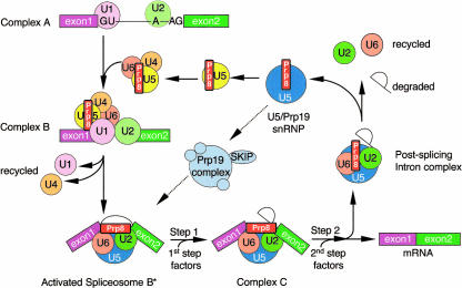
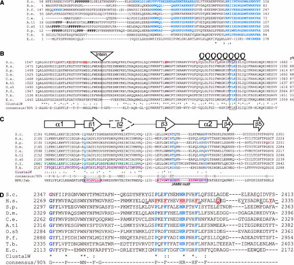
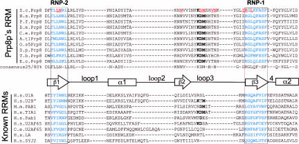
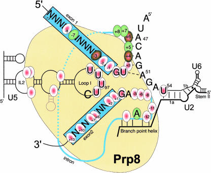
Pre-messenger RNA (pre-mRNA) splicing is a central step in gene expression. Lying between transcription and protein synthesis, pre-mRNA splicing removes sequences (introns) that would otherwise disrupt the coding potential of intron-containing transcripts. This process takes place in the nucleus, catalyzed by a large RNA-protein complex called the spliceosome. Prp8p, one of the largest and most highly conserved of nuclear proteins, occupies a central position in the catalytic core of the spliceosome, and has been implicated in several crucial molecular rearrangements that occur there. Recently, Prp8p has also come under the spotlight for its role in the inherited human disease, Retinitis Pigmentosa.Prp8 is unique, having no obvious homology to other proteins; however, using bioinformatical analysis we reveal the presence of a conserved RNA recognition motif (RRM), an MPN/JAB domain and a putative nuclear localization signal (NLS). Here, we review biochemical and genetical data, mostly related to the human and yeast proteins, that describe Prp8's central role within the spliceosome and its molecular interactions during spliceosome formation, as splicing proceeds, and in post-splicing complexes.


Figures

FIGURE 1.
FIGURE 1.
Schematic diagram showing the assembly and recycling of spliceosome components with respect to Prp8. The conserved 5′ and 3′ splice sites represent the U2 cis-spliceosomal GU and AG residues, respectively. Prp8 has been found in two different U5 complexes: the U5 snRNP (small, yellow) and the larger U5/Prp19 snRNP (large, blue). (Adapted with permission from Makarov et al. © 2002 AAAS [www.sciencemag.org].)
FIGURE 2.
FIGURE 2.
Maps showing regions of Prp8p against which antibodies have been generated. The regions recognized are represented as arrows below the antibody names. S. cerevisiae antibodies 8.1–8.4 were raised against lacZ–protein fusions (Lossky et al. 1987; Jackson et al. 1988) whereas all the others are raised against peptides. α-8.1 only precipitates yPrp8 in U5 snRNPs, whereas α-8.4 and α-8.6 are the only ones that precipitate yeast spliceosomes and co-complexes. The 70R antibody against hPrp8 (Luo et al. 1999) is noted as being superior to KO5 for immunoprecipitation. One other antibody to tbPrp8 is known (Lucke et al. 1997).
FIGURE 3.
FIGURE 3.
Alignments of 10 Prp8 orthologs showing four conserved motifs. Important residues are highlighted in bold blue type and known alleles underlined in red. (S.c.) Saccharomyces cerevisiae; (H.s.) Homo sapiens; (P.f.) Plasmodium falciparum; (D.m.) Drosophila melonagaster; (C.e.) Caenorhabditis elegans; (A.t1.) Arabidopsis thaliana chromosome 1; (O.s5) Oryza sativa chromosome 5; (S.p.) Schizosaccharomyces pombe; (T.b.) Trypanosoma buceii; (E.c.) Encephalitazoon cuniculii. Numbers refer to the amino acid residues. All sequences aligned using ClustalW and consensii are derived from 25 Prp8 orthologs using the ‘consensus’ program. (A) Alignment of the N-terminal regions. The polyproline tracts can be seen in the nonconserved N termini of some species. Potential bipartite NLS sequences are highlighted in bold blue type and are separated by 15 amino acids. In the case of S. cerevisiae an overlapping nuclear localization signal (NLS) sequence is underlined. In the cases of P. falciparum and E. cuniculi, a shorter SV40 type NLS and pat7 types are highlighted, respectively. The NLS sequences always occur within the first 500 N-terminal residues as predicted by PSORTII (Nakai and Horton 1999). (B) The highly conserved Prp8 domain 3.2. The position of the intein, present in four orthologs is indicated by the triangle. A potential protein kinase C phosphorylation site (T/S-x-R/K) that is conserved in all species is boxed and highlighted. This is central to the postulated coiled-coil domain represented above the text by the coil. (y%) The level of conservation compared with the yeast sequence. (C) The MPN/JAB domain found in Prp8. The mutations yprp8–28 and sprp8spp42–1 are shown in bold red underlined text. The representative MPN/JAB domain from NCBI’s domain database is shown at the bottom of the alignment. Two important MPN motif residues identified previously are boxed in plum coloured text (Maytal-Kivity et al. 2002). The five residues in the MPN structure that coordinate the zinc are highlighted in bold blue text. The secondary structure diagrams are shown above the alignment (from Tran et al. 2003). (D) The C terminus of Prp8 showing the single mutated residues altered in human Retinitis Pigmentosa (bold, red underlined type), the nonsense mutation (bold, red, and circled) and the residues where frameshift mutations occur (doublly underlined text). The temperature-sensitive yprp8–1 mutation affects the first glycine residue on the left-hand side of the alignment (bold plum).
FIGURE 4.
FIGURE 4.
Prp8 contains a conserved RNA recognition motif (RRM). Using a hidden Markov model with the HMMER package (Eddy 1998) against the human NCBI database, only RRM containing proteins were identified. (The model is available as Supplementary Data at http://www.ed.ac.uk/~jeanb/; Supplemental Fig. S1). The alignment between 10 different Prp8 sequences and nine known RRMs from the Interpro database is carried out using ClustalW or T-coffee in combination with Jalview (Thompson et al. 1994; Notredame et al. 2000). The conserved RNP-1 and RNP-2 motifs are boxed and highlighted in bold blue type. A number of mutations are known in yeast Prp8 that suppress brr2–1 and/or U4–cs1 mutations and these are highlighted by bold red underlined text. A unique feature of Prp8’s RRM is the conserved amino acid triplet KDM in the center of the domain. The consensus calculations are derived from 25 different Prp8p sequences from 23 species using ClustalW and the online “consensus” program. A stylized representation of the RRM secondary structure based on the U1A crystal structure is shown between the primary sequences.
FIGURE 5.
FIGURE 5.
A summary of the RNA residues that are known to cross-link to Prp8 in the context of the spliceosome. Cross-linked residues are circled, the exons are boxed, and the intron is represented by a thin blue line. (Red circles) Short-range cross-links using uridine and 4-thiouridine; (green circles) long-range cross-links using the reagent benzophenone. Where both cross-linking agents were used at the same site the circles colored red and green with a dotted outline. Extensive Prp8 cross-links with U5 snRNA are also represented as is the U6 residue 54. Both the U5 and U6 cross-links were only demonstrated in snRNPs. All numbering relates to S. cerevisiae.
FIGURE 6.
FIGURE 6.
Pictoral representations of (A) proteins shown to interact with yeast Prp8p, (B) gene expression coupled with pre-mRNA splicing, and (C) three purified Prp8-containing protein complexes involving the nuclear receptor activated promoter VDR (Zhang et al. 2003), the transcription elongation complex (Kameoka et al. 2004) and the spliceosome (Makarov et al. 2002). (Gray circle) RNA pol II; (rectangle and red oval) the spliceosome. The interchromatin granular clusters (IGC), or “speckles” as they are also known, store/assemble the RNAPII and splicing factors. Upon chromatin remodelling, the DNA becomes accessible, the RNAPII/spliceosome proteins assemble on the perichromatin fibrils (PF) and produce pre-mRNA. After removal of the intron the mRNA is exported through the nuclear pore (NP) for translation and RNAPII/spliceosome proteins are stored or reassembled in the IGCs.
FIGURE 7.
FIGURE 7.
A stylized diagram of yPrp8 mapping the known motifs and yeast mutations. The numbers along the length of Prp8 represent the amino acid numbers for human and yeast. On the left are the yeast proteins known to interact with yPrp8 aligned approximately with the yPrp8 minimal region of interaction. (Y2H) yeast two hybrid; (Ph.D) phage display; (…) protein–protein interactions. Down the center of the stylized Prp8p are the motifs and domains discussed in the text. On the right a conservation bar code shows only residues that are 100% conserved between nineteen different species. The two plasmodium sequences were omitted because they contain excessive insertion sequences. The similarity line-up was performed with Vector NTI (Alignment; Invitrogen). In the table on the right are the allele names and their relevant genotype/amino acid alteration. A simple coloured dots scheme highlights their phenotypes. Alleles have then been aligned against the amino acid conservation map of Prp8 using blue trapezoids and yellow highlighting. (prp28Δ*) prp28Δ only in the presence of yhc-1; (S.L.) synthetic lethal; (Pyr) pyrimidine tract recognition mutation; (x) alleles tested and no phenotype noted; (ts) temperature sensitive; (cs) cold sensitive. The lab that first reported the mutations is indicated by the lab head’s initials; references numbers refer to a list posted on Jean Beggs’ Web site (http://www.ed.ac.uk/~jeanb/). P.I. initials and names are: (DB) D. Brow; (TC) T-C Chang; (JB) J. Beggs; (CG) C. Guthrie; (MK) M. Konarska; (MKu) Martin Kupiec; (XF) X. Fu; (LJ) L. Johnston; (BS) B. Schwer; (DX) D. Xu; and (CI) C. Inglehearn.

Similar articles

Cited by

References

    1. Abovich, N. and Rosbash, M. 1997. Cross-intron bridging interactions in the yeast commitment complex are conserved in mammals. Cell 89: 403–412. - PubMed
    1. Achsel, T., Ahrens, K., Brahms, H., Teigelkamp, S., and Luhrmann, R. 1998. The human U5–220kD protein (hPrp8) forms a stable RNA-free complex with several U5-specific proteins, including an RNA unwindase, a homolog of ribosomal elongation factor EF-2, and a novel WD-40 protein. Mol. Cell. Biol. 18: 6756–6766. - PMC - PubMed
    1. Ajuh, P., Kuster, B., Panov, K., Zomerdijk, J.C., Mann, M., and Lamond, A.I. 2000. Functional analysis of the human CDC5L complex and identification of its components by mass spectrometry. EMBO J. 19: 6569–6581. - PMC - PubMed
    1. Albers, M., Diment, A., Muraru, M., Russell, C.S., and Beggs, J.D. 2003. Identification and characterization of Prp45p and Prp46p, essential pre-mRNA splicing factors. RNA 9: 138–150. - PMC - PubMed
    1. Alvi, R.K., Lund, M., and Okeefe, R.T. 2001. ATP-dependent interaction of yeast U5 snRNA loop 1 with the 5′ splice site. RNA 7: 1013–1023. - PMC - PubMed

Publication types