. 2005 May;11(5):646-56.
doi: 10.1261/rna.7198205.

Seawi--a sea urchin piwi/argonaute family member is a component of MT-RNP complexes

Affiliations

Seawi--a sea urchin piwi/argonaute family member is a component of MT-RNP complexes

Alexis J Rodriguez et al. RNA. 2005 May.

Abstract

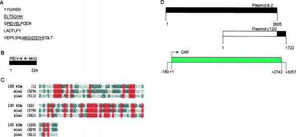
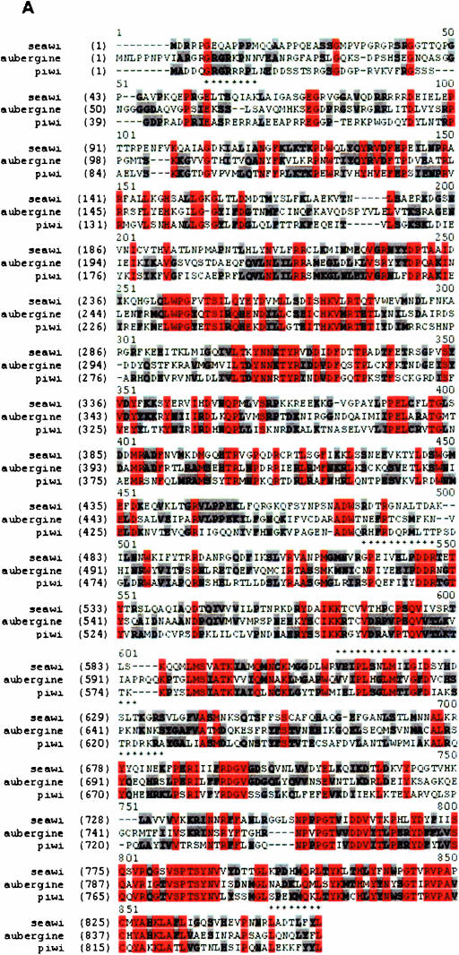
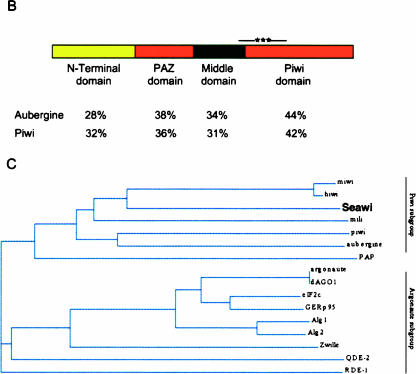
The piwi/argonaute family of proteins is involved in key developmental processes such as stem cell maintenance and axis specification through molecular mechanisms that may involve RNA silencing. Here we report on the cloning and characterization of the sea urchin piwi/argonaute family member seawi. Seawi is a major component of microtubule-ribonucleoprotein (MT-RNP) complexes isolated from two different species of sea urchin, Strongylocentrotus purpuratus and Paracentrotus lividus. Seawi co-isolates with purified ribosomes, cosediments with 80S ribosomes in sucrose density gradients, and binds microtubules. Seawi possesses the RNA binding motif common to piwi family members and binds P. lividus bep4 mRNA, a transcript that co-isolates with MT-RNP complexes and whose translation product has been shown to play a role in patterning the animal-vegetal axis. Indirect immunofluorescence studies localized seawi to the cortex of unfertilized eggs within granule-like particles, the mitotic spindle during cell division, and the small micromeres where its levels were enriched during the early cleavage stage. Lastly, we discuss how seawi, as a piwi/argonaute family member, may play a fundamentally important role in sea urchin animal-vegetal axis formation and stem cell maintenance.


Figures

FIGURE 1.
FIGURE 1.
Molecular cloning of seawi. (A) The five peptide microsequences obtained following proteolytic degradation of the 100-kDa protein. The underlined residues highlight the sequences used for synthesis of degenerate primers. (B) Schematic representation of the result of RT-PCR of total egg RNA with PEIV and MIIG primers with the orientation of the primers shown with arrows. (C) Alignment of the translated sequence of the 324-bp product with piwi and miwi generated using Vector NTI Suite 6.0 software (InforMax, Inc.) AlignX program. Residues identical in all three sequences are shown in red while residues identical in two of the sequences are shown in gray. (D) Schematic representation of the products from cDNA library screening. The black areas in plasmid 6-2 and plasmid c12/2 show the areas used to determine the consensus sequence of the 100-kDa protein. The bottom diagram shows key features of the consensus sequence.
FIGURE 2.
FIGURE 2.
Classification of the 100-kDa protein (seawi) as a member of the piwi family. (A) Alignment of the full-length sequence of the 100-kDa protein with piwi and aubergine. Residues identical in all three sequences are shown in red while residues identical in two of the sequences are shown in gray. The asterisks above the seawi sequence identify the five peptide microsequences. (B) Domain organization of seawi. The level of identity of each of the domains with the corresponding regions from piwi and aubergine are shown. The asterisks above the piwi domain within seawi identify the putative mRNA binding domain based on sequence homology with miwi’s mRNA binding sequence. (C) Phylogenetic analysis generated using Vector NTI Suite Version 6.0 software AlignX program. Note, seawi clusters most closely to hiwi and miwi.
FIGURE 2.
FIGURE 2.
Classification of the 100-kDa protein (seawi) as a member of the piwi family. (A) Alignment of the full-length sequence of the 100-kDa protein with piwi and aubergine. Residues identical in all three sequences are shown in red while residues identical in two of the sequences are shown in gray. The asterisks above the seawi sequence identify the five peptide microsequences. (B) Domain organization of seawi. The level of identity of each of the domains with the corresponding regions from piwi and aubergine are shown. The asterisks above the piwi domain within seawi identify the putative mRNA binding domain based on sequence homology with miwi’s mRNA binding sequence. (C) Phylogenetic analysis generated using Vector NTI Suite Version 6.0 software AlignX program. Note, seawi clusters most closely to hiwi and miwi.
FIGURE 3.
FIGURE 3.
Seawi is the 100-kDa protein of S. purpuratus MT-RNP complexes. (A) SDS-PAGE gel of MT-RNP complex proteins and purified seawi. (Lane 1) Isolated MT-RNP complex. (Lane 2) Purified seawi protein. (B) Characterization of seawi antisera. (Lane 1) Bio-Rad prestained high range SDS-PAGE standards (molecular weights are the same as those shown in A). (Lane 2) SDS-PAGE of total protein from S. purpuratus egg. (Lane 3) Immunoblot of total egg protein with anti-100-kDa protein polyclonal antiserum (4I4). (Lane 4) Immunoblot of total egg protein with preimmune serum for 4I4. (Lane 5) Immunoblot of total egg protein with anti-100-kDa MT-RNP complex protein polyclonal antiserum (A59). (Lane 6) Immunoblot of total egg protein with preimmune serum for A59. (C) Companion immunoblots to the SDS-PAGE of the MT-RNP complex (left lane) and purified seawi (right lane) shown in Panel A. Blots were probed using either anti-100-kDa protein polyclonal antiserum (4I4) or anti-100-kDa MT-RNP complex protein polyclonal antiserum (A59); only the relevant portions of the blots are shown. Each antiserum binds to both of the 100-kDa proteins, thus establishing their immuno-relatedness.
FIGURE 4.
FIGURE 4.
Seawi is a component of purified ribosome preparations. Coomassie blue stained gel of S. purpuratus MT-RNP complex and purified ribosome preparation are shown in lanes 2 and 3, respectively. Companion immunoblots (MT-RNP complex, lane 4; purified ribosome preparation, lane 5) were probed using antisera against the 100-kDa protein/seawi (A59), EMAP, α-tubulin, and a 40-kDa component of ribosomes (ribo). Molecular weight standards are shown in lane 1.
FIGURE 5.
FIGURE 5.
Sucrose density centrifugation analysis of S. purpuratus ribosome preparations. (A) Silver stained SDS-PAGE gel of the results of sucrose gradient centrifugation of purified egg ribosomes (lane L) with the corresponding regions of the companion immunoblots probed with A59 antiserum (seawi) and anti-40-kDa ribosomal protein (ribo). The position of the 80S ribosome is indicated, demonstrating that seawi and the 80S ribosome cosediment. The top of the gradient is at the left. (B) Silver stained SDS-PAGE gel of the results of sucrose gradient centrifugation of purified egg ribosomes following high salt treatment. The arrows indicate the positions of the dissociated 40S and 60S ribosomal subunits. Notice that seawi fails to cosediment with either of the ribosomal subunits under these conditions.
FIGURE 6.
FIGURE 6.
Comparison of S. purpuratus and P. lividus MT-RNP complexes. Coomassie blue stained SDS-PAGE gels of S. purpuratus (lane 1) and P. lividus (lane 2) MT-RNP complex preparation. (Lane 3) Result of an immunoblot of the P. lividus MT-RNP complex probed with anti-seawi antibodies (A59).
FIGURE 7.
FIGURE 7.
Seawi binds bep4 mRNAs. A Northwestern blot of purified LP54 (lane 1), purified seawi (lane 2), and purified fibronectin (lane 3) probed with full-length bep4 message. Notice that LP54 (previously shown to be an mRNA binding protein); (Montana et al. 1997) and seawi bind bep4 message while the control protein, fibronectin, does not.
FIGURE 8.
FIGURE 8.
Developmental expression profile of seawi. Levels of seawi message (red) and protein (blue) present at different stages of development were normalized to the amount of message or protein found in the unfertilized eggs. (inset) Representative immunoblot of total S. purpuratus protein probed with A59 antiserum. The following stages were investigated: unfertilized eggs (lane 0); 6 h, 4 divisions (lane 1); 12 h, 8 divisions (lane 2); 18 h, late blastula (lane 3); 23 h, primary mesenchyme ingression (lane 4); 31 h, gastrula (lane 5); 42 h, archenteron formation (lane 6); 52 h, prism (lane 7); 72 h, pluteus larva (lane 8).
FIGURE 9.
FIGURE 9.
Immunolocalization of seawi. (A) Laser scanning confocal micrograph of an unfertilized S. purpuratus egg stained with A59 antiserum. (B) Laser scanning confocal micrograph of a cortical preparation of an unfertilized egg stained with 4I4 antiserum. (C) Higher magnification of a laser scanning confocal micrograph of a cortical preparation of an unfertilized egg stained with 4I4 antiserum. (D) Laser scanning confocal micrograph of a first division S. purpuratus embryo stained with A59 antiserum. (E) Laser scanning confocal micrograph of an early cleavage stage S. purpuratus embryo stained with A59 antiserum. Notice the enrichment of seawi in the four micromeres present at the vegetal pole of the embryo. The bars in A,B,D,E are 10 μm, while the bar in C is 1 μm.

Similar articles

Cited by

References

    1. Angerer, L.M. and Angerer, R.C. 2000. Animal–vegetal axis patterning mechanisms in the early sea urchin embryo. Dev. Biol. 218: 1–12. - PubMed
    1. Aplin, A.E. and Juliano, R.L. 2001. Regulation of nucleocytoplasmic trafficking by cell adhesion receptors and the cytoskeleton. J. Cell Biol. 155: 187–191. - PMC - PubMed
    1. Barbarese, E., Koppel, D.E., Deutscher, M.P., Smith, C.L., Ainger, K., Morgan, F., and Carson, J.H. 1995. Protein translation components are colocalized in granules in oligodendrocytes. J. Cell Sci. 108: 2781–2790. - PubMed
    1. Bonder, E.M., Fishkind, D.J., Cotran, N.M., and Begg, D.A. 1989. The cortical actin-membrane cytoskeleton of unfertilized sea urchin eggs: Analysis of the spatial organization and relationship of filamentous actin, nonfilamentous actin, and egg spectrin. Dev. Biol. 134: 327–341. - PubMed
    1. Brandhorst, B.P. and Klein, W.H. 2002. Molecular patterning along the sea urchin animal–vegetal axis. Int. Rev. Cytol. 213: 183–232. - PubMed

Publication types

MeSH terms

LinkOut - more resources