. 2005 May;11(5):717-27.
doi: 10.1261/rna.2340405.

A role for eIF4E and eIF4E-transporter in targeting mRNPs to mammalian processing bodies

Affiliations

A role for eIF4E and eIF4E-transporter in targeting mRNPs to mammalian processing bodies

Maria Alexandra Andrei et al. RNA. 2005 May.

Abstract

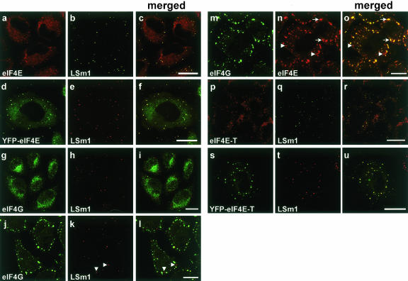
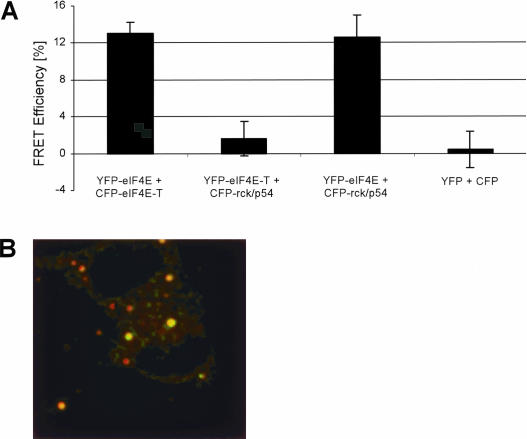
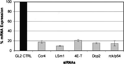
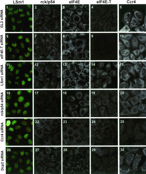
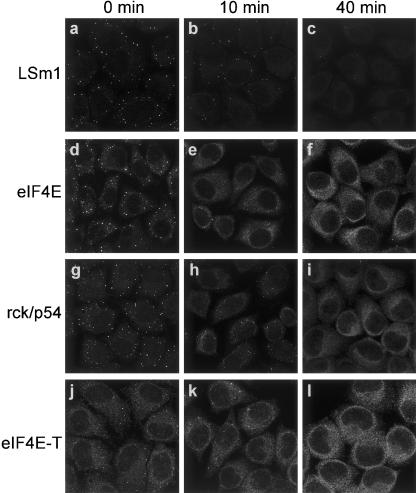
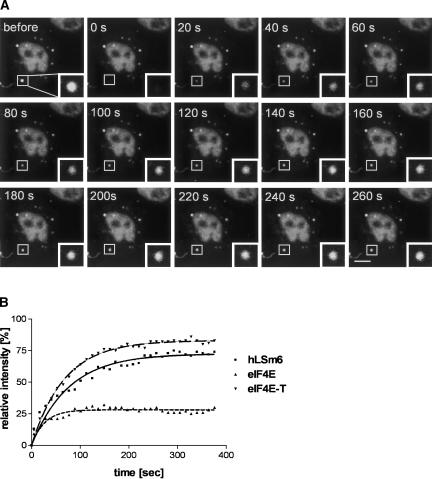
mRNP remodeling events required for the transition of an mRNA from active translation to degradation are currently poorly understood. We identified protein factors potentially involved in this transition, which are present in mammalian P bodies, cytoplasmic foci enriched in 5' --> 3' mRNA degrading enzymes. We demonstrate that human P bodies contain the cap-binding protein eIF4E and the related factor eIF4E-transporter (eIF4E-T), suggesting novel roles for these proteins in targeting mRNAs for 5' --> 3' degradation. Furthermore, fluorescence resonance energy transfer (FRET) studies indicate that eIF4E interacts with eIF4E-T and the putative DEAD box helicase rck/p54 in the P bodies in vivo. RNAi-mediated knockdowns revealed that a subset of P body factors, including eIF4E-T, LSm1, rck/p54, and Ccr4 are required for the accumulation of each other and eIF4E in P bodies. In addition, treatment of HeLa cells with cycloheximide, which inhibits translation, revealed that mRNA is also required for accumulation of mRNA degradation factors in P bodies. In contrast, knockdown of the decapping enzyme Dcp2, which initiates the actual 5' --> 3' mRNA degradation did not abolish P body formation, indicating it first functions after mRNPs have been targeted to these cytoplasmic foci. These data support a model in which mRNPs undergo several successive steps of remodeling and/or 3' trimming until their composition or structural organization promotes their accumulation in P bodies.


Figures

FIGURE 1.
FIGURE 1.
eIF4E and eIF4E-T colocalize with LSm1 in distinct cytoplasmic foci. HeLa SS6 cells were grown on coverslips, fixed, and stained with antibodies specific for eIF4E (monoclonal anti-eIF4E, Santa Cruz Biotechnology) (A,N), LSm1 (B,E,H,K,Q,T), eIF4G (G,J,M), or eIF4E-T (P). Alternatively, cells were transfected with plasmids encoding YFP-eIF4E (D) or YFP-eIF4E-T (S). Panels C,F,I,L,O,R,U show the merged picture of the proceeding two panels, with overlaying signals appearing yellow. In panels JO, cells were treated with 100 μM arsenite for 45 min at 37°C to induce stress granules (indicated by arrows). Cells were analyzed by confocal fluorescence microscopy. Scale bars = 10 μm.
FIGURE 2.
FIGURE 2.
eIF4E and eIF4E-T proteins interact directly in vivo, as demonstrated by FRET. (A) Bar chart representing the mean values of the apparent FRET efficiencies of protein pairs from several P bodies in different cells. Error bars indicate the standard deviation from the mean values.The protein pair CFP-eIFE-T/YFP-eIF4E was coexpressed (B). HeLa SS6 cells were fixed 16 h after transfection with 4% PFA and confocal images of both CFP and YFP channels were taken before and after photobleaching. The difference in post- and prebleach (of the YFP acceptor) intensity of the CFP-fluorescence was divided by the postbleach CFP-fluorescence. The ratio was calculated pixel by pixel, and the result is shown color coded. Green indicates a FRET efficiency of 15%, red indicates a FRET efficiency near 0 (no FRET). The image was contrast enhanced to also display the dim P bodies, in the prebleach donor image, which is the brightness channel of this image. The total size of this image corresponds to 56 μm in the sample.
FIGURE 3.
FIGURE 3.
Efficient knockdown of Ccr4, LSm1, eIF4E-T, Dcp2, and rck/p54 mRNA levels revealed by real-time RT-PCR. HeLa SS6 cells growing in six-well cell cultures dishes were transfected with either the GL2 control siRNA or with siRNAs against Ccr4, LSm1, eIF4E-T, Dcp2, and rck/p54 at a concentration of 120 nM. After 48 h cells were harvested and total RNA was extracted and incubated with RQ1 Dnase; 50 ng of total RNA was used in one-step RT-PCR (QuantiTec SYBR Green RT-PCR Kit). The graph shows the reduction in target mRNA levels calculated from the real-time RT-PCR data compared to mRNA levels from cells transfected with GL2 control siRNA. Error bars indicate the standard deviation from several independent experiments.
FIGURE 4.
FIGURE 4.
eIF4E-T, LSm1, rck/p54, and Ccr4, but not Dcp2, are required for the accumulation of each other in P bodies. HeLa cells were transfected with GL2 luciferase control (1–5), eIF4E-T (610), LSm1 (11–15), rck/p54 (16–20), Ccr4 (21–25), and Dcp2 (26–30) siRNA duplexes. Cells were immunostained with antibodies against LSm1 (panels 1,6,11,16,21,26), rck/p54 (panels 2,7,12,17,22,27), eIF4E (panels 3,8,13,18,23,28), eIF4E-T (panels 4,9,14,19,24,29), and Ccr4 (panels 5,10,15,20,25,30). Cells immunostained for LSm1 were counterstained with CybrGold showing cell nuclei (green). The panels show two-dimensional projections (Zeiss Software) of a series of confocal fluorescence images in order to obtain sharp images displaying all P bodies in every cell. To highlight the important structures, different magnifications were used for the various panels.
FIGURE 5.
FIGURE 5.
Effect of cycloheximide on the accumulation of LSm1, eIF4E, rck/p54, and eIF4E-T in P bodies. Cells were incubated with cycloheximide (20 μg/mL) and fixed at 0 min, 10 min, and 40 min after addition of inhibitor. Cells were immunostained with antibodies against LSm1 (a–c), eIF4E (d–f), rck/p54 (g–i), and eIF4E-T (j–l). The panels represent two-dimensional projections (Zeiss Software) of a series of confocal fluorescence images (see legend to Fig. 2 ▶).
FIGURE 6.
FIGURE 6.
Fluorescence recovery after photobleaching indicates that P bodies are highly dynamic in nature. HeLa cells were transfected with plasmids encoding YFP-LSm6, YFP-eIF4E, or YFP-eIF4E-T. (A) In an YFP-LSm6-transfected cell a P body (see rectangle) was recorded (“before”), bleached (“0 s”), and images were collected every 20 sec during the course of recovery. (B) For each time point fluorescence intensity within the region of interest was measured and plotted in the graph shown relative to the intensity before bleaching. From each data set, fluorescence recovery curves were calculated using nonlinear regression. Fitting was carried out by the Marquardt method (GraphPad Software). Scale bar = 10 μm.

Similar articles

Cited by

References

    1. Bashkirov, V.I., Scherthan, H., Solinger, J.A., Buerstedde, J.M., and Heyer, W.D. 1997. A mouse cytoplasmic exoribonuclease (mXRN1p) with preference for G4 tetraplex substrates. J. Cell. Biol. 136: 761–773. - PMC - PubMed
    1. Bastiaens, P.I. and Jovin, T.M. 1998. Fluorescence energy transfer microscopy (FRET). In Cell biology: A laboratory handbook (ed. J.E. Celis), Vol. III, pp. 136–146. Academic Press, New York.
    1. Chen, J., Chiang, Y.C., and Denis, C.L. 2002. CCR4, a 3′-5′ poly(A) RNA and ssDNA exonuclease, is the catalytic component of the cytoplasmic deadenylase. EMBO J. 21: 1414–1426. - PMC - PubMed
    1. Coller, J.M., Tucker, M., Sheth, U., Valencia-Sanchez, M.A., and Parker, R. 2001. The DEAD box helicase, Dhh1p, functions in mRNA decapping and interacts with both the decapping and deadenylase complexes. RNA 7: 1717–1727. - PMC - PubMed
    1. Cougot, N., Babajko, S., and Seraphin, B. 2004. Cytoplasmic foci are sites of mRNA decay in human cells. J. Cell Biol. 165: 31–40. - PMC - PubMed

Publication types

MeSH terms

LinkOut - more resources