. 2005 May;14(5):1201-13.
doi: 10.1110/ps.041256705.

Structural consequences of the familial amyotrophic lateral sclerosis SOD1 mutant His46Arg

Affiliations

Structural consequences of the familial amyotrophic lateral sclerosis SOD1 mutant His46Arg

Svetlana Antonyuk et al. Protein Sci. 2005 May.

Abstract

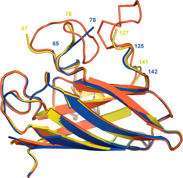
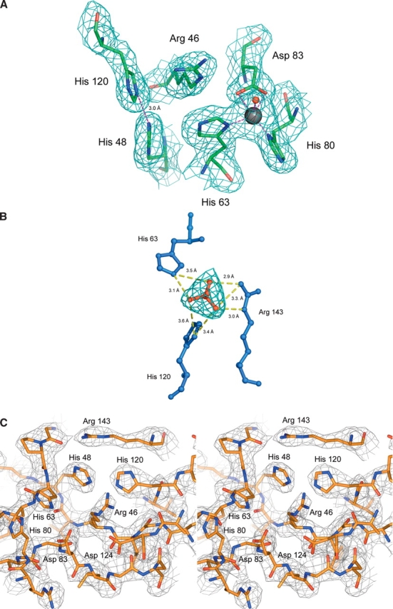
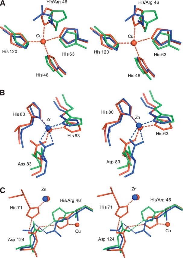
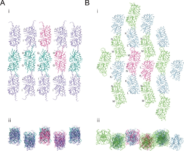
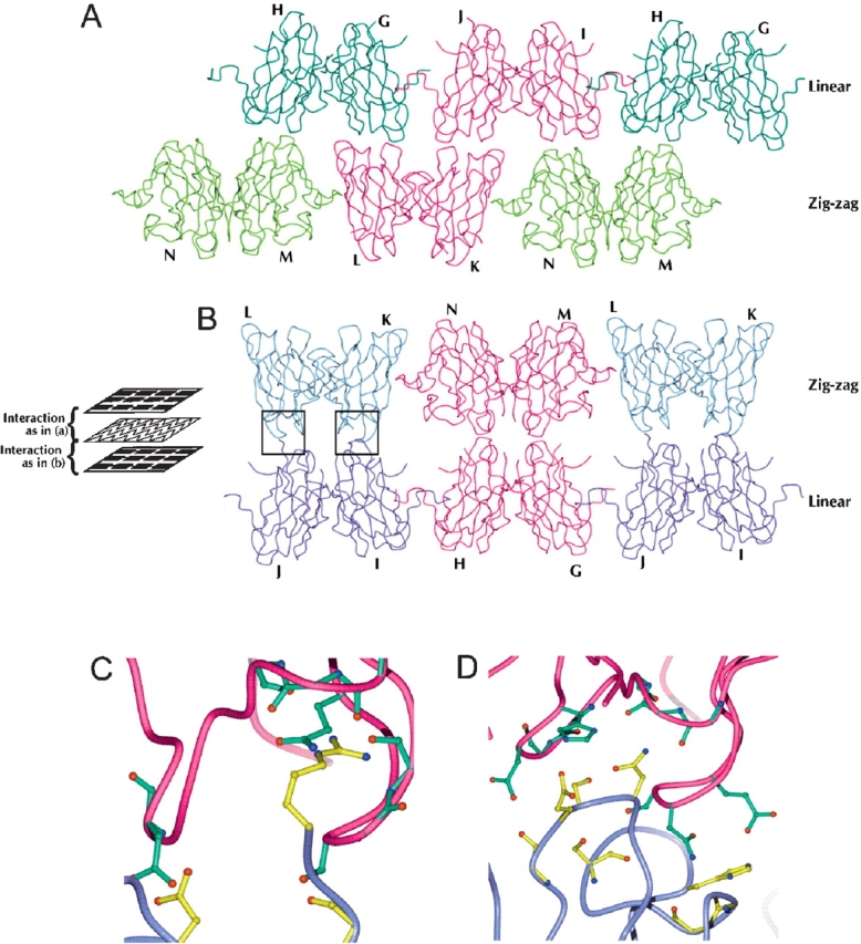
The His46Arg (H46R) mutant of human copper-zinc superoxide dismutase (SOD1) is associated with an unusual, slowly progressing form of familial amyotrophic lateral sclerosis (FALS). Here we describe in detail the crystal structures of pathogenic H46R SOD1 in the Zn-loaded (Zn-H46R) and metal-free (apo-H46R) forms. The Zn-H46R structure demonstrates a novel zinc coordination that involves only three of the usual four liganding residues, His 63, His 80, and Asp 83 together with a water molecule. In addition, the Asp 124 "secondary bridge" between the copper- and zinc-binding sites is disrupted, and the "electrostatic loop" and "zinc loop" elements are largely disordered. The apo-H46R structure exhibits partial disorder in the electrostatic and zinc loop elements in three of the four dimers in the asymmetric unit, while the fourth has ordered loops due to crystal packing interactions. In both structures, nonnative SOD1-SOD1 interactions lead to the formation of higher-order filamentous arrays. The disordered loop elements may increase the likelihood of protein aggregation in vivo, either with other H46R molecules or with other critical cellular components. Importantly, the binding of zinc is not sufficient to prevent the formation of nonnative interactions between pathogenic H46R molecules. The increased tendency to aggregate, even in the presence of Zn, arising from the loss of the secondary bridge is consistent with the observation of an increased abundance of hyaline inclusions in spinal motor neurons and supporting cells in H46R SOD1 transgenic rats.


Figures

Figure 1.
Figure 1.
Superposition of a monomer of holo-wtSOD1 (red), apo-H46R (yellow), and Zn-H46R (blue). The apo-H46R monomer (G) is disordered in residues 68–77 (zinc loop) and 128–140 (electrostatic loop). The Zn-H46R structure is disordered in residues 66–77 and 126–141. The largest structural shifts between Zn-H46R and wtSOD1 occur in residues 118–125 and 78–80. In the latter case, the first residue after the break in the electron density Glu 78 is >7 Å away from its position in wtSOD1. In the case of the truncations of the model at residues 65 and 142, structural changes are limited to the final modeled residue. Superposition of the Zn-H46R dimers onto the best-defined dimer of the native hSOD structure yields a root mean square deviation (RMSD) for Cα atoms of 0.97 Å (not including those residues absent from the Zn-H46R model). Comparison of apo-H46R with the wild-type SOD1 dimer yields a RMSD of ~0.35 Å for all atoms shared between the two structures. The dimer interface is not altered by the H46R mutation in either structure.
Figure 2.
Figure 2.
(A) 2Fobs-Fcalc electron density maps, contoured at 1σ, for the copper and zinc sites of Zn-H46R. The copper site is vacant, and the mutated residue Arg 46 is well-defined. The zinc site is fully occupied, and the zinc ion is ligated by only three residues, His 63, His 80, and Asp 83. The usual fourth zinc ligand, His 71, is disordered and not present in the electron density. A water molecule (shown as a red sphere) acts as an additional weak ligand some 2.4 Å from Zn. The copper ligands His 48 and His 120 are stabilized by a 3 Å hydrogen bond between their Nɛ2 atoms. (B) Binding of sulfate near to the vacant copper site in Zn-H46R. Sulfate forms hydrogen bonds to Arg 143 and to the copper ligands His 63 and His 120, thus stabilizing the site in the absence of metal (see Table 3). (C) Stereo image of the structure and electron density around the altered active site of the copper and zinc sites of apo-H46R SOD1 monomer G. The 2.5 Å σA-weighted electron density, with coefficients 2mFo - DFc, is contoured at 1σ. Note the lack of metal ion electron density and novel hydrogen bonding between His 48 and His 120 of the SOD1 copper site and His 63 and His 80 of the SOD1 zinc site. Asp 83 and Asp 124 (the secondary bridging residue) are within hydrogen bonding distance of mutated Arg 46, further stabilizing the site. The fourth zinc ligand, His 71, exhibits no electron density and therefore was not modeled in this monomer.
Figure 3.
Figure 3.
(A) Stereo figures showing the superposition of Zn-H46R (blue), wtSOD1 (red), and apo wtSOD1 (green) for the copper site. The Nη1 atom of Arg 46 occupies a position 2.8 Å from the position occupied by copper in the native hSOD structure, while the Cɛ atom resides some 2.9 Å away. The new conformation of His 120 in Zn-H46R allows the copper-ligating Nɛ2 atoms of histidines 48 and 120 to form a ~3 Å hydrogen bond. His 48 remains in a very similar conformation in the mutant and wtSOD1 structures. This preserved conformation is likely to be a consequence of the hydrogen bond between its Nδ1 atom and the O atom of Gly 61. The side chain of His 120 is shifted, occupying a position slightly closer to the vacant copper position (Nɛ2 to copper distance would be ~1.8 Å). (B) The zinc site. In Zn-H46R zinc is ligated by only three protein residues where Asp 83 acts as a bidentate ligand. His 71 is absent in Zn-H46R and is omitted from this figure for clarity, as is the water ligand observed in the Zn-H46R structure. (C) The secondary bridge. In wtSOD1, residue Asp 124 forms a hydrogen-bonded link between the copper ligand His 46 and the zinc ligand His 71. Asp 124 Oδ2 forms a strong (~2.6 Å) hydrogen bond to His46 Nɛ2, while its Oδ1 atom bonds to His71 Nɛ2. In addition, the Oδ1 atom is close to the backbone nitrogen atoms of residues 125 (3.1 Å) and 126 (2.8 Å) of the electrostatic loop, providing additional stabilization of the structure. In Zn-H46R, the mutant residue 46 maintains hydrogen bonding to Asp 124, but residue His 71 is disordered and the bridge is broken. In apo-wtSOD1, in the absence of metals His 46 hydrogen bonds to Asp 124, which adopts a different rotamer to that present in wtSOD1.
Figure 4.
Figure 4.
(A) End-on view of a plane of helical filaments in the Zn-H46R structure. The crystallographic asymmetric unit consists of a pair of SOD1 dimers (boxed). (B) Surface representation showing the interface between monomers of Zn-H46R arising from adjacent helical filaments. The interacting residues Asp 11 and Lys 36 are shown in a ball-and stick representation. A further interaction involves reciprocal interactions of Pro 13.
Figure 5.
Figure 5.
Sheets of filaments in the apo-H46R structure. (A) Linear filament sheet: planar view (i) and end-on view (ii). Filaments are arranged anti-parallel to each other with the axis of propagation determined by head-to-head interactions between the altered conformation electrostatic loop elements of the G/H (dark green) and I/J (purple) dimers. The G/H (bottom right) and I/J (top left) components of the asymmetric unit are colored dark pink. The sheet is connected by tenuous water-mediated side-to-side interaction between linear filaments between monomers G and H. (B) Zigzag packing interaction sheet: planar view (i) and end-on view (ii). These interactions are arranged anti-parallel to each other with the axis of propagation determined by head-to-head interactions between the altered conformation electrostatic loop elements of the K/L (blue) and M/N (light green) dimers. The K/L (right) and M/N (left) components of the asymmetric unit are in dark pink. The sheet packing is completed by side-to-side interactions between two of the zigzag filament subunits, chains L and N.
Figure 6.
Figure 6.
Interactions between apo-H46R filament planes. The asymmetric unit consists of dimers G/H, I/J, L/K, and M/N. The linear filament and zigzag interaction planes lie parallel to each other in space as layers. The apo-H46R molecules of each plane make interactions of two types between these layers perpendicular to the filament planes: between the bottom surface of the linear plane and the top surface of the zigzag plane (A) and between the bottom surface of the zigzag plane and the top surface of the linear plane (B) (see schematic). Dimers G/H (dark green) and I/J (purple) form the linear filament layer, and L/K (blue) and M/N (light green) dimers form the zigzag layer. The components of the asymmetric unit are in dark pink. There are three sites of interaction between the four molecules within the asymmetric unit and consequently between the two classes of filament: (1) a few hydrogen-bond, water-mediated, and van der Waals interactions between the region of chain M near Asp 109 and the area around the same residue in chain G; (2) a similar interaction involving residues around the Asp 109 residues in monomers N and H; and (3) reciprocal van der Waals interactions only between residues 12–13 and 37–39 of chains K and J. The most extensive of the interactions between the linear and zigzag sheets occurs between monomers L and J and K and I, which form a total of 30 side-chain–to–side-chain and side-chain–to–main-chain hydrogen bonds through their respective areas adjacent to and including part of the zinc loop (C,D). Another interaction involves reciprocal hydrogen bonds between the β-strand connecting loops (residues 13–15, 36–39, and 91–92) of monomers L and G. The third interaction occurs through the same area of chains I and N but is very tenuous, consisting of only one electrostatic interaction between residues Asp 92 and Lys 36 and a few van der Waals interactions. (C) Closeup of hydrogen bonding interactions between monomers L and J at the zigzag and linear plane interface (left boxed region in B). (D) Closeup of hydrogen bonding interactions between monomers K and I at the zigzag and linear plane interface (right boxed region in B). (C,D) The K/L dimer is shown in dark pink, and the I/J dimer is in purple. Residues involved in the interaction have either their side chain or backbone modeled in detail, green (K/L) or yellow (I/J), depending on which atoms of the residue are directly involved in hydrogen bonding.

References

    1. Aoki, M., Ogasawara, M., Matsubara, Y., Narisawa, K., Nakamura, S., Itoyama, Y., and Abe, K. 1994. Familial amyotrophic-lateral-sclerosis (ALS) in Japan associated with H46R mutation in Cu/Zn superoxide-dismutase gene: A possible new subtype of familial ALS. J. Neurol. Sci. 126 77–83. - PubMed
    1. Aoki, M., Abe, K., and Itoyama, Y. 1998. Molecular analyses of the Cu/Zn superoxide dismutase gene in patients with familial amyotrophic lateral sclerosis (ALS) in Japan. Cell. Mol. Neurobiol. 18 639–647. - PMC - PubMed
    1. Arisato, T.O.R., Arata, H., Abe, K., Fukada, K., Sakoda, S., Shimizu, A., Qin, X.H., Izumo, S., Osame, M., and Nakagawa, M. 2003. Clinical and pathological studies of familial amyotrophic lateral sclerosis (FALS) with SOD1 H46R mutation in large Japanese families. Acta Neuropathol. 106 561–568. - PubMed
    1. Banci, L., Felli, I.C., and Kummerle, R. 2002. Direct detection of hydrogen bonds in monomeric superoxide dismutase: Biological implications. Biochemistry 41 2913–2920. - PubMed
    1. Bernstein, F.C., Koetzle, T.F., Williams, G.J.B., Meyer Jr., F.M., Brice, M.D., Rodgers, J.R., Kennard, O., Shimanouchi, T., and Tasumi, M. 1977. The Protein Data Bank: A computer based archival file for macromolecular structures. J. Neurol. Biol. 112 535–542. - PubMed

Publication types