Review
. 2005 Apr;115(4):798-812.
doi: 10.1172/JCI24826.

Immunopathogenesis and therapy of cutaneous T cell lymphoma

Affiliations
Review

Immunopathogenesis and therapy of cutaneous T cell lymphoma

Ellen J Kim et al. J Clin Invest. 2005 Apr.

Erratum in

  • J Clin Invest. 2007 Mar;117(3):836

Abstract

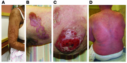
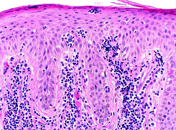
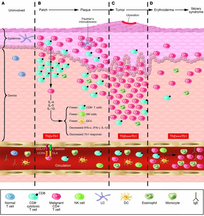
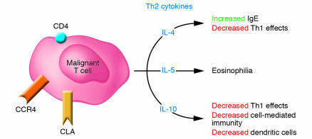
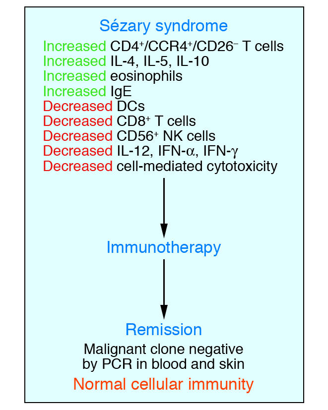
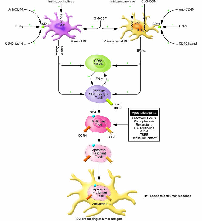
Cutaneous T cell lymphomas (CTCLs) are a heterogenous group of lymphoproliferative disorders caused by clonally derived, skin-invasive T cells. Mycosis fungoides (MF) and Sezary syndrome (SS) are the most common types of CTCLs and are characterized by malignant CD4(+)/CLA(+)/CCR4(+) T cells that also lack the usual T cell surface markers CD7 and/or CD26. As MF/SS advances, the clonal dominance of the malignant cells results in the expression of predominantly Th2 cytokines, progressive immune dysregulation in patients, and further tumor cell growth. This review summarizes recent insights into the pathogenesis and immunobiology of MF/SS and how these have shaped current therapeutic approaches, in particular the growing emphasis on enhancement of host antitumor immune responses as the key to successful therapy.


Figures

Figure 1
Figure 1
Cutaneous lesions of MF and SS. (A) Hypopigmented patches of MF on the proximal arm. Patches can be pink, red, hypopigmented, or hyperpigmented and are often scaly. (B) Two plaques near the axilla. MF lesions can have annular and gyrate configurations. (C) A tumor on the arm. Tumors frequently ulcerate. (D) Erythroderma in a patient with SS; more than 80% of his body surface area is affected with confluent erythematous scaly patches.
Figure 2
Figure 2
Histopathology of classic MF. This skin biopsy specimen demonstrates an atypical lymphocytic infilitrate going up into the epidermis (epidermotropism) in the absence of epidermal edema (spongiosis). The collection of atypical lymphocytes surrounding a Langerhans cell is a Pautrier microabcess, the hallmark of classic MF.
Figure 3
Figure 3
The skin microenvironment in MF progression. (A) Normal skin showing resident Langerhans cells in the epidermis and skin-homing T cells in the dermis and circulation. (B) Patch and plaque MF in which the CD4+ malignant T cells home to the epidermis and collect around Langerhans cells. Of note, in these stages, the epidermal and dermal infiltrate frequently have abundant CD8+ T cells as part of the host immune response. (C) Tumor MF in which the tumor occupies the dermis and subcutaneous tissue and is comprised of primarily malignant T cells and few CD8+ T cells. (D) Erythrodermic MF and SS with detectable circulating malignant T cells that elaborate Th2 cytokines that affect CD8+ T cell, NK cell, and DC numbers and function, and consequently, the host immune response.
Figure 4
Figure 4
Consequences of malignant T cell cytokine production. In MF/SS, the malignant T cell (CD4+/CLA+/CCR4+) produces the cytokines IL-4, IL-5, and IL-10 that result in a Th2 predominance and subsequent multiple abnormalities in cellular immunity.
Figure 5
Figure 5
Elimination of the malignant T cell clone during immunotherapy leads to a restoration of a normal immune response. Studies of numerous patients with SS have demonstrated that induction of complete remission with clearing of the malignant T cell clone during multimodality immunotherapy leads to a restoration of normal host immune function.
Figure 6
Figure 6
Multimodality strategy for enhancing the antitumor response using immunotherapeutics. A multimodality approach encompasses the activation of multiple arms of the immune response through the use of agents to activate DCs, CTLs (CD8+), and NK cells (CD56+). GM-CSF may enhance the numbers of DCs while agents that enhance CD40 expression (IFN-γ) and activation (CD40 ligand; activating anti-CD40 antibody) and TLR ligands (CpG-ODNs; imidazoquinolines) lead to DC cytokine production and to enhanced DC processing of apoptotic malignant T cells. Cytokines produced by DCs as well as exogenously administered cytokines augment CD8+ T cell and possibly NK cell cytolytic activity against the tumor cells. RAR-specific retinoids stimulate DC and CD8+ T cell cytokine production. Proapoptotic agents, including bexarotene, RAR-specific retinoids, PUVA, photopheresis, topical chemotherapy, total skin electron beam irradiation (TSEB), and denileukin diftitox, can assist in the development of an antitumor immune response by reducing the overall tumor burden and by providing a source of apoptotic malignant cells and tumor antigens for uptake by DCs.
Figure 7
Figure 7
Inhibiting the effects of regulatory T cells. By inhibiting soluble factors produced by regulatory T cells, preventing CTLA-4 engagement of CD80 or CD86, and inducing apoptosis of regulatory T cells by agents such as denileukin diftitox, the overall effects of regulatory T cells on the immune response can be diminished.

References

    1. Girardi M, Heald PW, Wilson LD. The pathogenesis of mycosis fungoides. N. Engl. J. Med. 2004;350:1978–1988. - PubMed
    1. Kazakov DV, Burg G, Kempf W. Clinicopathological spectrum of mycosis fungoides. J. Eur. Acad. Dermatol. Venereol. 2004;18:397–415. - PubMed
    1. Willemze, R., et al. 2005. WHO-EORTC classification for cutaneous lymphomas. Blood. doi:10.1182/blood-2004-09-3502. - PubMed
    1. Leboit, P.E., and McCalmont, T.H. 1997. Cutaneous lymphomas and leukemias. In Lever’s histopathology of the skin. D. Elder, C. Jaworsky, and B. Johnson, editors. Lippincott-Raven. Philadelphia, Pennsylvania, USA. 820 pp.
    1. Kim YH, et al. Long-term outcome of 525 patients with mycosis fungoides and Sezary syndrome: clinical prognostic factors and risk for disease progression. Arch. Dermatol. 2003;139:857–866. - PubMed