. 2005 Apr;115(4):870-8.
doi: 10.1172/JCI21386.

Induction of dermal-epidermal separation in mice by passive transfer of antibodies specific to type VII collagen

Affiliations

Induction of dermal-epidermal separation in mice by passive transfer of antibodies specific to type VII collagen

Cassian Sitaru et al. J Clin Invest. 2005 Apr.

Abstract

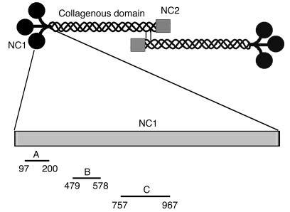
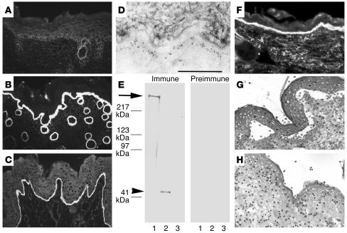
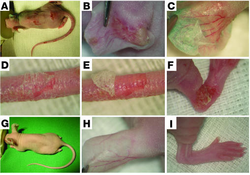
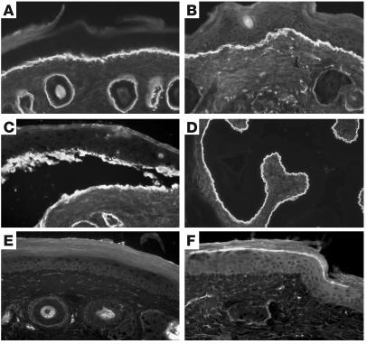
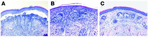
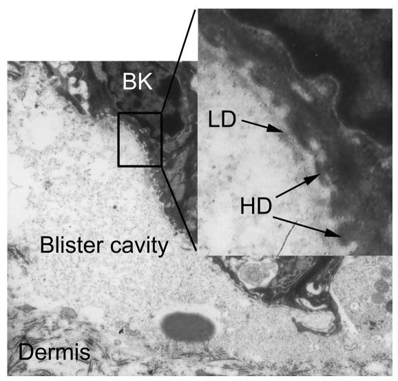
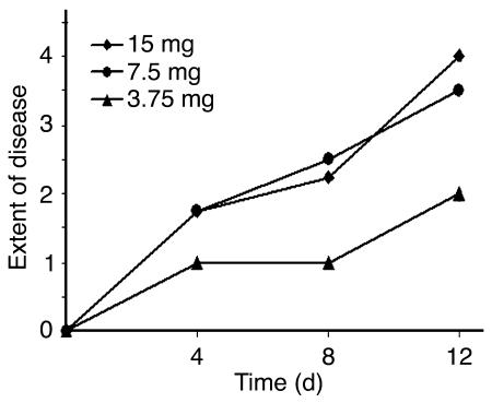
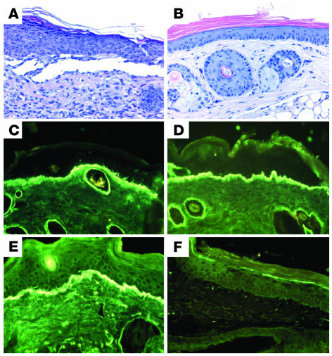
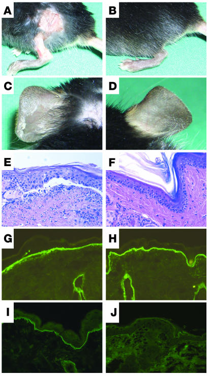
Epidermolysis bullosa acquisita (EBA) is a subepidermal blistering disorder associated with tissue-bound and circulating autoantibodies specific to type VII collagen, a major constituent of the dermal-epidermal junction. Previous attempts to transfer the disease by injection of patient autoantibodies into mice have been unsuccessful. To study the pathogenic relevance of antibodies specific to type VII collagen in vivo, we generated and characterized rabbit antibodies specific to a murine form of this antigen and passively transferred them into adult nude, BALB/c, and C57BL/6 mice. Immune rabbit IgG bound to the lamina densa of murine skin and immunoblotted type VII collagen. Mice injected with purified IgG specific to type VII collagen, in contrast to control mice, developed subepidermal skin blisters, reproducing the human disease at the clinical, histological, electron microscopical, and immunopathological levels. Titers of rabbit IgG in the serum of mice correlated with the extent of the disease. F(ab')(2) fragments of rabbit IgG specific to type VII collagen were not pathogenic. When injected into C5-deficient mice, antibodies specific to type VII collagen failed to induce the disease, whereas C5-sufficient mice were susceptible to blister induction. This animal model for EBA should facilitate further dissection of the pathogenesis of this disease and development of new therapeutic strategies.


Figures

Figure 1
Figure 1
Schematic organization of human type VII collagen and cDNA constructs generated for this study for expressing recombinant peptides of the NC1 domain. Type VII collagen is composed of 3 identical α chains, each consisting of a central triple helical collagenous domain, flanked by a large amino-terminal noncollagenous domain (NC1) and a smaller carboxy-terminal noncollagenous domain (NC2). Two molecules form antiparallel tail-to-tail dimers stabilized by disulfide bonding through a carboxy-terminal overlap between NC2 domains. Three fragments of murine type VII collagen cDNA were cloned in pGEX-6P-1 and expressed in E. coli. Amino acid residue numbers are shown below the fragments.
Figure 2
Figure 2
Serum IgG from rabbit SA2954 binds to the DEJ, recognizes type VII collagen, and activates complement and leukocytes in vitro. IF microscopy of rabbit SA9254 serum on frozen skin sections shows no specific staining before immunization (A); in contrast, after immunization with type VII collagen, IgG binds to the DEJ of mouse (B) and human (C) skin (magnification, ×250). (D) By immunoelectron microscopy, IgG purified from immune rabbit serum binds to the lamina densa of mouse skin. Scale bar: 0.5 μm. (E) Extract of murine dermis (lane 1) and equimolar amounts of GST-mCOL7C (lane 2) and GST (lane 3) were separated by gradient 4–20% SDS-PAGE and immunoblotted with immune and preimmune rabbit serum preadsorbed against GST. IgG from immune serum, but not preimmune serum, binds to both full-length cell-derived (arrow) and recombinant fragment C (arrowhead) of type VII collagen. (F) Frozen murine skin sections were incubated with immune rabbit serum and subsequently with fresh serum as a source of complement. Bound murine C3 was visualized at the DEJ by FITC-labeled antibody (magnification, ×250). When incubated with frozen sections of human skin in the presence of human leukocytes, IgG from the immune (G), but not preimmune (H) rabbit–induced dermal-epidermal separation was observed (magnification, ×400).
Figure 3
Figure 3
IgG to murine type VII collagen induces cutaneous lesions in adult mice. (A) Extensive skin lesions, including blisters, erosions, and epidermal detachment, developed in a BALB/cnu/nu mouse receiving, over a period of 10 days, 5 injections of IgG, each containing 7.5 mg of IgG, from a rabbit immunized against murine type VII collagen. Injections into the back of the mouse induced (B) spontaneous blisters on the left ear and, upon tangential pressure, (C) epidermal detachment on the right ear. (D) Epidermal detachment was also elicited on tail skin, and (E) the epidermis could subsequently be easily lifted up from the dermis. (F) Erosions covered by crusts on the hind limb. (GI) A control mouse challenged with the same dose of normal rabbit IgG showed no skin alterations.
Figure 4
Figure 4
Rabbit IgG specific to type VII collagen binds to the basement membrane and fixes complement in vivo. IF microscopy, performed on frozen sections of a perilesional mouse skin biopsy (A and B) and murine esophagus (D) reveals linear deposition of rabbit IgG (A and D) and murine C3 (B) at the basement membrane in a diseased mouse. (C) IF analysis of lesional skin from a diseased mouse shows rabbit IgG deposits on both epidermal and dermal sides of the cleavage. No deposits of rabbit IgG (E) or murine C3 (F) are detected at the DEJ of a mouse injected with control rabbit IgG (magnification, ×250).
Figure 5
Figure 5
By light microscopy, antibodies specific to type VII collagen cause subepidermal splits. (A) Histologic examination of skin biopsies from a diseased mouse reveals extensive subepidermal cleavage (magnification, ×200). (B) The inflammatory infiltrate is dominated by neutrophils (magnification, ×400). (C) No specific histologic alterations are found in the skin of a mouse treated with control IgG (magnification, ×200).
Figure 6
Figure 6
Experimentally induced splits localize below the lamina densa. Electron microscopic examination of a lesional skin biopsy from a diseased mouse demonstrates that the blister roof contains the lamina densa (LD) bordered by basal keratinocytes (BK) with hemidesmosomes (HD) (arrows). Dermal connective tissue represents the blister floor (magnification, ×11,000; inset: magnification, ×44,000).
Figure 7
Figure 7
The extent of cutaneous disease in the mice depends on the injected amount of immune rabbit IgG. The extent of disease was scored as described in Methods and in Table 2. Means of individual clinical scores (n = 4) are shown before the first injection as well as 4, 8, and 12 days later. Significantly more extensive disease was induced in mice injected with 7.5 mg or 15 mg of immune IgG compared with mice treated with 3.75 mg of IgG.
Figure 8
Figure 8
Dermal-epidermal separation is dependent on the Fc portion of antibodies specific to type VII collagen. (A) Subepidermal blister in a skin biopsy from a mouse receiving, over a period of 12 days, 6 injections of IgG, each containing 7.5 mg of intact IgG from immune rabbit serum. (B) In contrast, no histological changes in a mouse injected with F(ab′)2 fragments generated by pepsin digestion of pathogenic rabbit IgG (H&E, magnification, ×200). IF analysis of perilesional skin from a diseased mouse revealed positive staining using both anti-Fab (C) and anti-Fc (E) FITC-conjugated antibodies. In a mouse injected with F(ab′)2 fragments, staining was observed with the Fab- (D) but not with the Fc-specific (F) conjugate (×400).
Figure 9
Figure 9
C5-deficient mice are resistant to the induction of cutaneous disease by antibodies specific to type VII collagen. Erosions on back and leg and, upon tangential pressure, epidermal detachment on the ear in a C5-sufficient (A and C) but not in a C5-deficient (B and D) mouse. Mice received 5 injections, each containing 15 mg of IgG from immune rabbit serum, over a period of 10 days. Histologic analysis of murine skin revealed subepidermal cleavage and a neutrophil-rich inflammatory infiltrate in C5-sufficient mice (E) but no histological changes in C5-deficient mice (F) (magnification, ×200). IF analysis of mouse skin showed deposition of rabbit IgG in mice both deficient (G) and sufficient (H) in C5. Deposits of membrane attack complex along the dermal-epidermal junction were only observed in C5-sufficient (I) and not in C5-deficient (J) mice (magnification, ×400).

Comment in

Similar articles

Cited by

References

    1. Rose NR, Bona C. Defining criteria for autoimmune diseases (Witebsky’s postulates revisited) Immunol. Today. 1993;14:426–430. - PubMed
    1. Schmidt E, Zillikens D. Autoimmune and inherited subepidermal blistering diseases: advances in the clinic and the laboratory. Adv. Dermatol. 2000;16:113–157. - PubMed
    1. Woodley DT, et al. Identification of the skin basement-membrane autoantigen in epidermolysis bullosa acquisita. N. Engl. J. Med. 1984;310:1007–1013. - PubMed
    1. Stanley JR, Rubinstein N, Klaus-Kovtun V. Epidermolysis bullosa acquisita antigen is synthesized by both human keratinocytes and human dermal fibroblasts. J. Invest. Dermatol. 1985;85:542–545. - PubMed
    1. Woodley DT, et al. Epidermolysis bullosa acquisita antigen is the globular carboxyl terminus of type VII procollagen. J. Clin. Invest. 1988;81:683–687. - PMC - PubMed

Publication types

MeSH terms