. 2005 Apr;115(4):879-87.
doi: 10.1172/JCI23977.

Synergy between a plasminogen cascade and MMP-9 in autoimmune disease

Affiliations

Synergy between a plasminogen cascade and MMP-9 in autoimmune disease

Zhi Liu et al. J Clin Invest. 2005 Apr.

Abstract

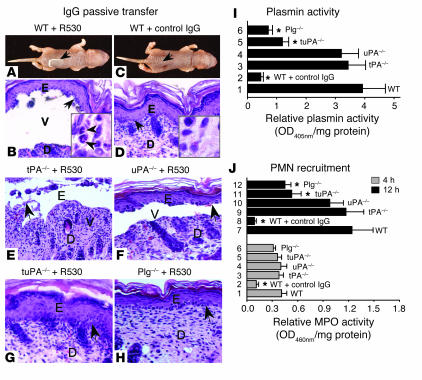
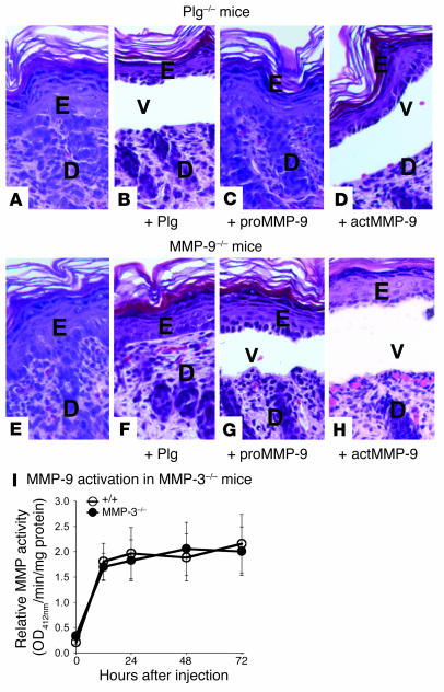
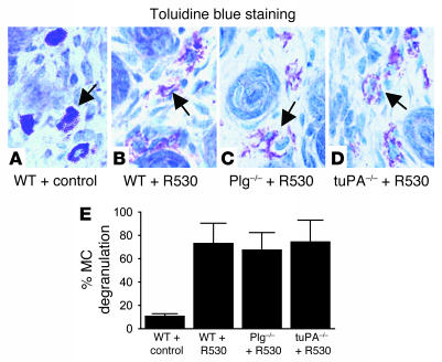
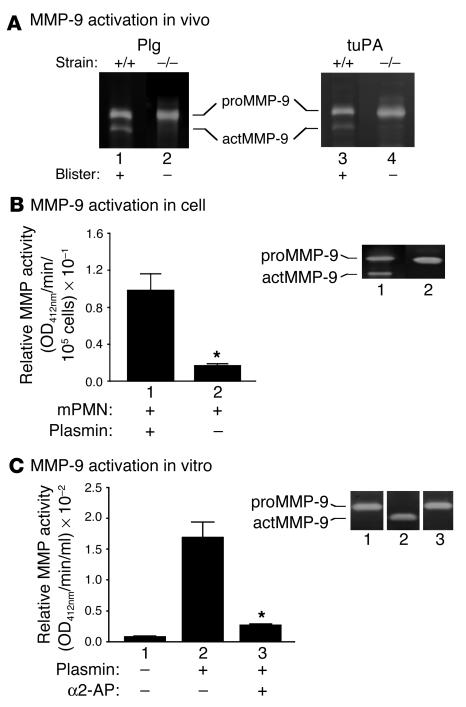
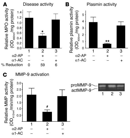
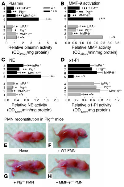
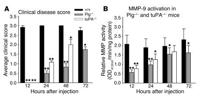
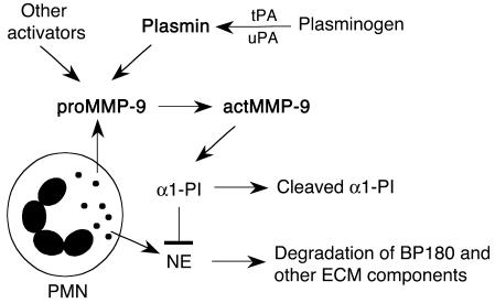
Extracellular proteolysis by the plasminogen/plasmin (Plg/plasmin) system and MMPs is required for tissue injury in autoimmune and inflammatory diseases. We demonstrate that a Plg cascade synergizes with MMP-9/gelatinase B in vivo during dermal-epidermal separation in an experimental model of bullous pemphigoid (BP), an autoimmune disease. BP was induced in mice by antibodies to the hemidesmosomal antigen BP180. Mice deficient in MMP-9 were resistant to experimental BP, while mice deficient in Plg and both tissue Plg activator (tPA) and urokinase Plg activator (uPA) showed delayed and less intense blister formation induced by antibodies to BP180. Plg-deficient mice reconstituted locally with Plg or the active form of MMP-9 (actMMP-9), but not the proenzyme form of MMP-9 (proMMP-9), developed BP. In contrast, proMMP-9 or actMMP-9, but not Plg, reconstituted susceptibility of MMP-9-deficient mice to the skin disease. In addition, MMP-3-deficient mice injected with pathogenic IgG developed the same degree of BP and expressed levels of actMMP-9 in the skin similar to those of WT controls. Thus, the Plg/plasmin system is epistatic to MMP-9 activation and subsequent dermal-epidermal separation in BP.


Figures

Figure 1
Figure 1
The Plg/plasmin system is required for experimental BP. WT mice and mice deficient in different components of the Plg/plasmin system were injected i.d. with pathogenic anti-mBP180 IgG (R530) or control IgG and examined 12 hours later. (AH) WT (A and B), tPA–/– (E), and uPA–/– (F), but not tuPA–/– (G) or Plg–/– (H) mice injected with pathogenic IgG developed subepidermal blisters. WT injected with control IgG showed no disease (C and D). Arrows indicate sites of basal keratinocytes. E, epidermis; D, dermis; V, blister vesicle. Magnification, ×200. Higher magnifications of H&E staining sections demonstrate infiltrating neutrophils in the dermis (insets). Arrowheads indicate neutrophils. Magnification, ×920. (I) Plasmin chromogenic assay showed significantly higher levels of plasmin activity in the lesional skin of WT (bar 1), tPA–/– (bar 3), and uPA–/– (bar 4) mice as compared with the nonlesional skin of control (bar 2), tuPA–/– (bar 5), and Plg–/– (bar 6) mice. Data shown are the mean ± SEM. n = 9 for each group; *P < 0.001 versus WT. (J) MPO activity assay at 12 hours after injection (black bars) showed significantly higher levels of PMN recruitment in the lesional skin of WT (bar 7), tPA–/– (bar 9), and uPA–/– (bar 10) mice as compared with tuPA–/– (bar 11) and Plg–/– (bar 12) mice. At 4 hours after injection (gray bars), all mice had similar numbers of infiltrating neutrophils. n = 9 for each group; *P < 0.001 versus WT.
Figure 2
Figure 2
Plasmin generation is upstream of MMP-9 activation. (AD) actMMP-9 reconstitution restores BP in Plg–/– mice. (A) Pathogenic anti-mBP180 IgG (R530, i.d. injection, 2.64 mg/g body weight) induced subepidermal blistering in neonatal Plg–/– mice reconstituted with mouse Plg (5 μg/g body weight) (B) and actMMP-9 (2.5 μg/g body weight) (D) but not proMMP-9 (2.5 μg/g body weight) (C) and PBS control (A). (EH) Plg reconstitution failed to restore BP in MMP-9–/– mice. Pathogenic anti-mBP180 IgG (i.d. injection, 2.64 mg/g body weight) induced subepidermal blistering in neonatal MMP-9–/– mice reconstituted with the same amount of proMMP-9 (G) and actMMP-9 (H) but not mouse Plg (F) or PBS control (E). Magnification, ×200. (I) Like +/+ mice, MMP-3–/– mice developed extensive clinical and histological blisters (Table 1) and compatible levels of MMP activity at 12-, 24-, 48-, and 72-hour time points as determined by MMP colorimetric assay (mean ± SEM).
Figure 3
Figure 3
MC activation in Plg- and tuPA-deficient mice. WT, Plg–/–, and tuPA–/– mice were injected i.d. with pathogenic IgG (R530; 2.64 mg/g body weight). At 2 hours after injection, when MC degranulation reached the peak level, skin sections were stained with toluidine blue solution. (AD) Toluidine blue staining showed similar degrees of MC degranulation in pathogenic IgG-injected WT and deficient mice. (E) The MCs in the dermis were counted and classified as degranulated (more than 10% of the granules exhibiting fusion or discharge) or normal (see Methods). There was no significant difference in MC degranulation among these mice (mean ± SEM). Arrows indicate MCs.
Figure 4
Figure 4
MMP-9 activation by plasmin in vivo and in vitro. (A) To identify MMP-9 activation in the lesional skin of experimental BP, neonatal Plg+/+, Plg–/–, tuPA+/+, and tuPA–/– mice were injected i.d. with pathogenic anti-mBP180 IgG. Skin samples were obtained at 4 hours after IgG injection, and protein extracts (30 μg/lane) were analyzed by gelatin zymography. Both proMMP-9 and actMMP-9 were seen in lesional skin samples of IgG-injected +/+ mice (lanes 1 and 3). In contrast, no actMMP-9 was found in skin samples of pathogenic IgG-injected Plg–/– (lane 2) and tuPA (lane 4) mice. (B) To show MMP-9 activation in a cell system, mouse neutrophils (mPMN; 1 × 10⁵) were stimulated by pathogenic anti-mBP180 IgG (5 μg/ml) plus mBP180 antigen (5 μg/ml) at 37°C for 1 hour. The supernatant was then incubated with plasmin (2 μg/ml) at 37°C for 16 hours. proMMP-9 released by neutrophils was activated by plasmin as shown by gelatin zymography (lane 1) and MMP colorimetric assay (bar 1). n = 6; *P < 0.001. (C) To show MMP-9 activation in vitro, the recombinant mouse proMMP-9 (1 μg) was incubated with plasmin (1 μg) in 100 μl of reaction buffer at 37°C for 6 hours and assayed by gelatin zymography and MMP colorimetric assay. Plasmin converted proMMP-9 to actMMP-9 (lane 2 and bar 2), and the activation was blocked by the plasmin inhibitor α2-AP (lane 3 and bar 3). n = 6; *P < 0.001.
Figure 5
Figure 5
Blocking plasmin activity abolishes experimental BP. To demonstrate that α2-AP treatment blocks plasmin activity and subsequent MMP-9 activation and blistering, WT mice were injected i.d. with pathogenic anti-mBP180 IgG (2.64 mg/g body weight) followed by local administration of PBS, the plasmin inhibitor α2-AP, or the neutrophil cathepsin G inhibitor α1-AC. Mice were examined at 12 hours after IgG injection. (A) Pathogenic anti-mBP180 IgG induced high levels of neutrophil infiltration and subepidermal blisters in +/+ mice (bar 1) and mice treated with α1-AC (bar 3) but not in mice treated with α2-AP (bar 2). n = 9; *P < 0.01. (B) Plasmin activity assay showed a significant reduction of tissue plasmin activity in the α2-AP–treated mice (bar 2) as compared with that in the PBS-treated (bar 1) and α1-AC–treated mice (bar 3). n = 8 for each group; **P < 0.001. (C) Zymography assay detected an actMMP-9 band in the skin of mice treated with PBS (lane 1) and α1-AC (lane 3) but not in the skin of mice treated with α2-AP (lane 2). MMP colorimetric assay revealed a significant reduction of active MMP levels in the skin samples of α2-AP–treated mice (bar 2) as compared with those in the lesional skin samples of PBS- (bar 1) and α1-AC–treated mice (bar 3). n = 8; #P < 0.05.
Figure 6
Figure 6
Functional relationship between plasmin, MMP-9, NE, and α1-PI in experimental BP. +/+, MMP-9–/–, Plg–/–, and tuPA–/– mice (n = 6) were injected with anti-mBP180 IgG and examined 4 hours and 12 hours later. (A) Plasmin activity assay showed similar levels of tissue plasmin activity in +/+ and MMP-9–/– mice at 4 hours but significantly higher levels of tissue plasmin activity in the lesional skin of +/+ (bar 5) mice compared with MMP-9–/– (bar 6), Plg–/– (bar 7), and tuPA–/– (bar 8) mice at 12 hours. (B) MMP colorimetric assay revealed increased levels of MMP activity in lesional skin of +/+ mice (bars 1 and 5) as compared with those in nonlesional skin of MMP-9–/– (bars 2 and 6), Plg–/– (bars 3 and 7), and tuPA–/– (bars 4 and 8) mice. (C) NE activity was significantly higher in the lesional skin of +/+ mice (bars 1 and 5) relative to MMP-9–/– (bars 2 and 6), Plg–/– (bars 3 and 7), and tuPA–/– (bars 4 and 8) mice. (D) NE inhibition assay showed a significantly reduced level of α1-PI in the lesional skin of +/+ mice (bars 1 and 5) as compared with the skin of MMP-9–/– (bars 2 and 6), Plg–/– (bars 3 and 7), and tuPA–/– (bars 4 and 8) mice. *P < 0.05 and **P < 0.001 for paired samples. (EH) Plg–/– mice, when reconstituted locally with 5 × 10⁵ neutrophils from +/+ (F), Plg–/– (G), or MMP-9–/– (H) mice, developed BP blisters 12 hours after pathogenic IgG injection. n = 6.
Figure 7
Figure 7
Plasmin-independent activation of MMP-9 in experimental BP. +/+, Plg–/–, and tuPA–/– mice (n = 6) were injected i.d. with pathogenic IgG (2.64 mg/g body weight) and examined at 12, 24, 48, and 72 hours after injection. Note that all tuPA–/– mice died by 72 hours. (A) At 12 hours, only +/+ mice developed clinical and histological blisters. After 24 hours, all mice showed clinical blistering, but disease severity (mean disease score ± SEM) in Plg–/– and tuPA–/– mice was significantly lower than in +/+ mice. (B) MMP colorimetric assay showed a significantly higher MMP activity in +/+ mice than in Plg–/– and tuPA–/– mice at all time points. *P < 0.05; **P < 0.001.
Figure 8
Figure 8
Working model of MMP-9 activation in experimental BP. Experimental BP is initiated by pathogenic anti-BP180 and depends on complement activation, MC degranulation, and neutrophil infiltration. Infiltrating neutrophils (PMNs) upon activation release NE, proMMP-9, and other proteinases. In the early stages of blistering, proMMP-9 is mainly activated by plasmin, which is generated from Plg by tPA and/or uPA. Plasmin and other activators of MMP-9 activate proMMP-9. actMMP-9 cleaves α1-PI to release NE. Unchecked NE degrades BP180 and other ECM components, resulting in dermal-epidermal separation.

Comment in

References

    1. Werb Z. ECM and cell surface proteolysis: regulating cellular ecology. Cell. 1997;91:439–442. - PubMed
    1. Collen D. The plasminogen (fibrinolytic) system. Thromb. Haemost. 1999;82:259–270. - PubMed
    1. Werb Z, Mainardi CL, Vater CA, Harris ED., Jr Endogenous activation of latent collagenase by rheumatoid synovial cells. Evidence for a role for plasminogen activator. N. Engl. J. Med. 1977;296:1017–1023. - PubMed
    1. Woessner, J.F., and Nagase, H. 2000. Matrix metalloproteinases and TIMPs. Oxford University Press. Oxford, United Kingdom. 240 pp.
    1. Stanley, J.R. 1999. Bullous pemphigoid. In Fitzpatrick’s dermatology in general medicine. I.M. Freedberg et al., editors. McGraw-Hill. New York, New York, USA. 666–671.

Publication types

MeSH terms