. 2005 Apr;115(4):888-99.
doi: 10.1172/JCI24185.

Genetic and functional characterization of human pemphigus vulgaris monoclonal autoantibodies isolated by phage display

Affiliations

Genetic and functional characterization of human pemphigus vulgaris monoclonal autoantibodies isolated by phage display

Aimee S Payne et al. J Clin Invest. 2005 Apr.

Abstract

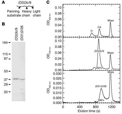
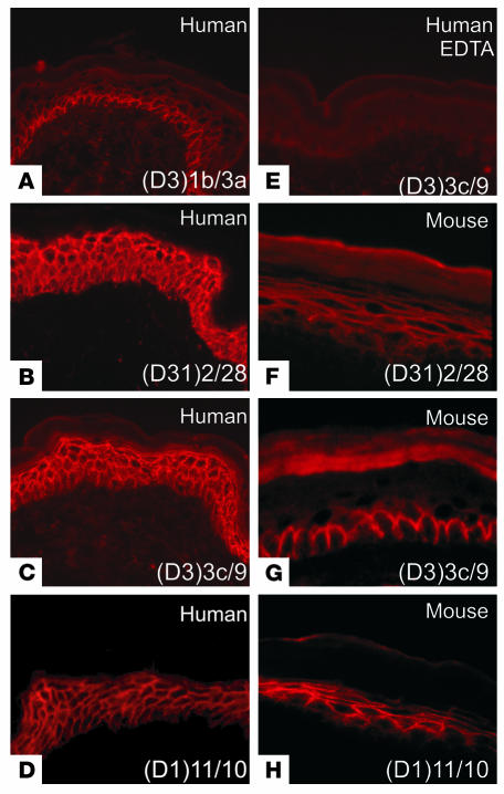
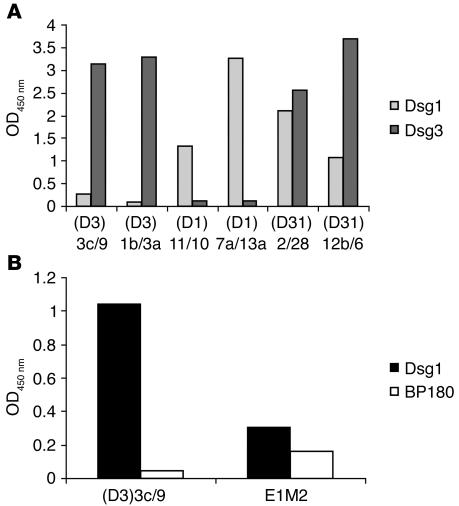
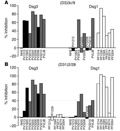
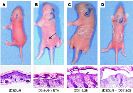
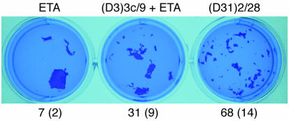
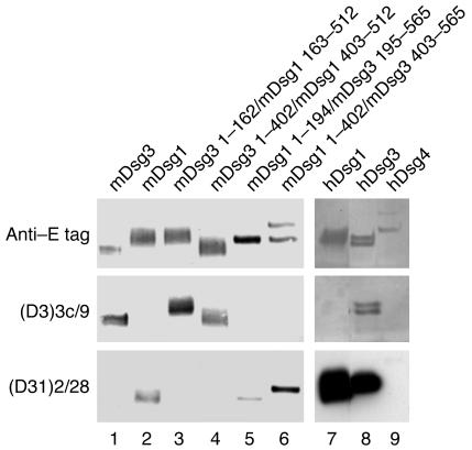
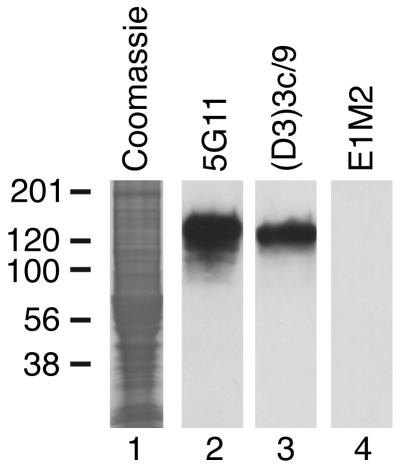
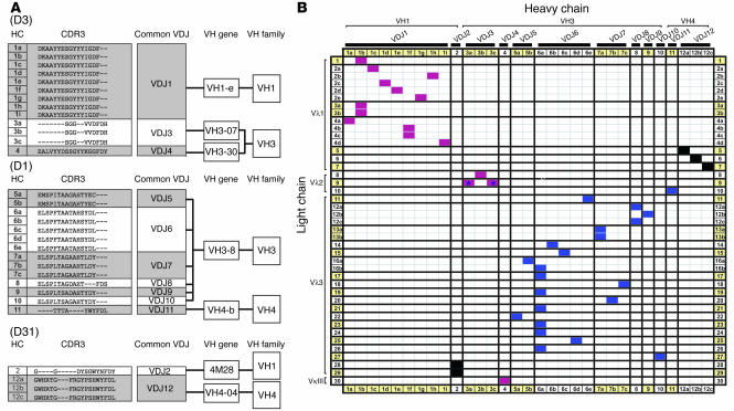
Pemphigus is a life-threatening blistering disorder of the skin and mucous membranes caused by pathogenic autoantibodies to desmosomal adhesion proteins desmoglein 3 (Dsg3) and Dsg1. Mechanisms of antibody pathogenicity are difficult to characterize using polyclonal patient sera. Using antibody phage display, we have isolated repertoires of human anti-Dsg mAbs as single-chain variable-region fragments (scFvs) from a patient with active mucocutaneous pemphigus vulgaris. ScFv mAbs demonstrated binding to Dsg3 or Dsg1 alone, or both Dsg3 and Dsg1. Inhibition ELISA showed that the epitopes defined by these scFvs are blocked by autoantibodies from multiple pemphigus patients. Injection of scFvs into neonatal mice identified 2 pathogenic scFvs that caused blisters histologically similar to those observed in pemphigus patients. Similarly, these 2 scFvs, but not others, induced cell sheet dissociation of cultured human keratinocytes, indicating that both pathogenic and nonpathogenic antibodies were isolated. Genetic analysis of these mAbs showed restricted patterns of heavy and light chain gene usage, which were distinct for scFvs with different desmoglein-binding specificities. Detailed characterization of these pemphigus mAbs should lead to a better understanding of the immunopathogenesis of disease and to more specifically targeted therapeutic approaches.


Figures

Figure 1
Figure 1
Isolation of monovalent scFv mAbs. (A) ScFv nomenclature. (D3), (D1), or (D31) indicates the antigens used to select anti-Dsg mAbs from the scFv phage display library (Dsg3, Dsg1, or both, respectively). This is followed by a unique heavy chain and light chain nucleotide sequence designation. For mAbs that share the same clonal origin (same VDJ rearrangement) but differ in sequence because of somatic hypermutation, lower-case letters indicate unique members of a given clone (see Discussion and legend to Figure 9). (B) Soluble scFv mAbs were purified by nickel-chelation chromatography (see Methods). Two representative scFvs are shown by Coomassie blue staining after SDS-PAGE. (C) Gel-filtration HPLC demonstrates that scFvs are primarily monomeric in solution. Transferrin (Tf, 75 kDa) and carbonic anhydrase (CA, 30 kDa) served as protein standards, with 0.1 M Mops as a marker of the total volume of the column. The percentage of monomeric protein is shown, calculated from the area under the curve.
Figure 2
Figure 2
IIF of scFv mAbs on human and mouse epidermis. The ScFv mAb used as primary antibody for staining human (AE) and mouse (FH) epidermis is indicated in each panel. Magnification, ×400.
Figure 3
Figure 3
Characterization of scFv mAb desmoglein-binding specificity by ELISA. (A) Patterns of the binding of selected scFv mAb clones to Dsg1 and Dsg3. (D3)3c/9 was isolated from a library panned on Dsg3 but showed quantifiable specificity for Dsg1, as shown in B. Similar results were obtained for mAb (D3)3a/9 (data not shown).
Figure 4
Figure 4
Epitopes recognized by PV scFv mAbs are blocked by sera from multiple pemphigus patients. (A) (D3)3c/9. Left: Inhibition of Dsg3 binding by all 8 PV sera. PVLIB is the plasma of the mucocutaneous PV patient from whom the phage display library was made. Black bars represent PV sera containing anti-Dsg3 antibodies only; gray bars indicate PV sera containing antibodies with both anti-Dsg3 and anti-Dsg1 activity [PV(3+1) sera]. Right: Inhibition of Dsg1 binding by 3 of 5 PV(3+1) sera and 6 of 6 PF sera (white bars). (B) (D31)2/28. Left: Inhibition of Dsg3 binding by 7 of 8 PV sera and 0 of 6 PF sera. Right: Inhibition of Dsg1 binding by 4 of 5 PV(3+1) sera and 5 of 6 PF sera. Inhibition of greater than 20% was considered positive. NHS, normal human sera.
Figure 5
Figure 5
Anti-Dsg scFv mAbs are pathogenic in neonatal mice. (A) Injection of (D3)3c/9 alone into neonatal mice does not cause gross blistering; histologic examination (lower panel) supports this result. (B) Injection of (D3)3c/9 with low-dose ETA demonstrates suprabasilar blistering. (C) Injection of (D31)2/28 alone demonstrates superficial blistering in the granular layer of the epidermis. (D) Coinjection of (D3)3c/9 and (D31)2/28 demonstrates suprabasilar blistering. Magnification, ×400.
Figure 6
Figure 6
ScFv mAbs cause dissociation of cultured human epidermal keratinocytes. Cultured human keratinocytes were incubated with ETA with or without scFvs and then treated with dispase to release cell monolayers. Released cell sheets were exposed to mechanical shear stress to evaluate intercellular adhesion. The total number of cell sheet fragments for each treatment is shown as a mean (SD).
Figure 7
Figure 7
Epitope mapping of (D3)3c/9 and (D31)2/28 against mouse and human desmogleins. Wild-type and domain-swapped extracellular domains of mDsg1 and mDsg3 (lanes 1–6) or wild-type extracellular domains of human desmogleins (hDsgs) 1, 3, and 4 (lanes 7–9) were produced in baculovirus and immunoprecipitated with scFv mAbs. Desmoglein proteins bound by human scFv mAbs were detected by immunoblot analysis using antibody against E tag, which was engineered onto the carboxyterminal domain of recombinant desmoglein molecules. The top panel shows the immunoblot of the recombinant and chimeric desmogleins detected with anti–E tag. The bottom 2 panels show the results of the immunoprecipitation-immunoblotting studies.
Figure 8
Figure 8
PV scFv mAbs recognize primarily but not exclusively conformational epitopes on desmogleins. Immunoblot analysis of human keratinocyte extract using (D3)3c/9. Lane 1, Coomassie blue–stained human keratinocyte extract. Lane 2, murine anti-Dsg3 mAb 5G11. Lane 3, (D3)3c/9. Lane 4, E1M2 (control anti–human red blood cell scFv).
Figure 9
Figure 9
Heavy and light chain restriction of Dsg-panned scFv mAbs. (A) Dendrogram and CDR3 alignment of (D3), (D1), and (D31) heavy chain sequences. Heavy chain (HC) sequence analysis of 63 randomly selected mAbs from the PV autoantibody repertoire identified 12 different VDJ families (indicated as VDJ1–VDJ12). Each VDJ family shares a common B cell precursor, defined by a common CDR3 amino acid sequence. The presence of a letter suffix in the heavy chain indicates the presence of somatic mutations in the variable region outside the CDR3, which reflect genetic diversification of the original parental B cell clone. The VH gene family and gene segment usage is indicated for each of the 31 unique heavy chain sequences. (B) Unique pemphigus mAbs show restricted and nonoverlapping usage of heavy and light chain gene segments. The horizontal axis represents unique heavy chains, and the vertical axis represents the unique λ and κ light chains (based on nucleic acid sequence). The heavy chains are restricted into 12 VDJ groupings, whereas the light chain repertoire comprises 30 groupings, defined by a unique light chain junctional region. Of the 26, 24, and 13 randomly screened D3, D1, and D31 mAbs, 16, 22, and 5 unique heavy and light chain combinations, respectively, were identified (represented within the matrix as magenta, blue, and black boxes, respectively). In most cases, antibodies panned against a particular desmoglein bound only that desmoglein substrate. However, 2 mAbs, (D3)3a/9 and (D3)3c/9, although panned only against Dsg3, also weakly bound Dsg1 (indicated by asterisks).

Comment in

References

    1. Payne AS, Hanakawa Y, Amagai M, Stanley JR. Desmosomes and disease: pemphigus and bullous impetigo. Curr. Opin. Cell Biol. 2004;16:536–543. - PubMed
    1. Ishii K, et al. Characterization of autoantibodies in pemphigus using antigen-specific enzyme-linked immunosorbent assays with baculovirus-expressed recombinant desmogleins. J. Immunol. 1997;159:2010–2017. - PubMed
    1. Ding X, et al. Mucosal and mucocutaneous (generalized) pemphigus vulgaris show distinct autoantibody profiles. J. Invest. Dermatol. 1997;109:592–596. - PubMed
    1. Amagai M, Tsunoda K, Zillikens D, Nagai T, Nishikawa T. The clinical phenotype of pemphigus is defined by the anti-desmoglein autoantibody profile. J. Am. Acad. Dermatol. 1999;40:167–170. - PubMed
    1. Miyagawa S, et al. Late development of antidesmoglein 1 antibodies in pemphigus vulgaris: correlation with disease progression. Br. J. Dermatol. 1999;141:1084–1087. - PubMed

Publication types

MeSH terms