. 2005 Apr;115(4):1021-9.
doi: 10.1172/JCI22486.

Ikaros integrates endocrine and immune system development

Affiliations

Ikaros integrates endocrine and immune system development

Shereen Ezzat et al. J Clin Invest. 2005 Apr.

Abstract

Ikaros transcription factors are essential regulators of lymphopoiesis and the development of the immune system. We now show that Ikaros is expressed in hormone-producing pituitary corticomelanotroph cells, where it binds the proopiomelanocortin promoter and regulates endogenous gene expression. Loss of Ikaros in vivo results in contraction of the pituitary corticomelanotroph population, reduced circulating adrenocorticotrophic hormone levels, and adrenal glucocorticoid insufficiency. While hemopoietic reconstitution failed to correct this hormonal deficit, the phenotype of reduced body weight and diminished survival was rescued by systemic glucocorticoid-hormone administration. Given the established immunomodulatory properties of glucocorticoid hormones, these findings reveal a novel role for Ikaros in orchestrating immune-endocrine development and function.


Figures

Figure 1
Figure 1
Identification of Ikaros and Eos expression in pituitary corticotroph cells. (A) Northern blotting of polyA RNA from pituitary corticomelanotroph AtT20 pituitary cells using Ikaros cDNA (top) identifies a doublet transcript of 2.7 and 2.5 kb, consistent with Ik1 and Ik2 mRNA expression. This doublet comigrates with that from pro–B lymphocyte–derived RNA. Eos mRNA expression (middle) is detected at much lower levels in AtT20 cells. The GAPDH loading control is shown immediately below. (B) Western blotting using specific antibodies against Ikaros (left) identifies a 52- and 50-kDa doublet that comigrates with that from lymphocytes. Detection with anti-Eos antibody (right) identifies the predicted 57-kDa product in AtT20 cells. The corresponding β-actin loading controls are shown immediately below. (C) Detection of Ikaros by EMSA of a DNA fragment including the Ikaros consensus binding motif, which binds nuclear extracts (NE) from pituitary corticomelanotroph AtT20 similar to those from pro-B lymphocytes. DNA-protein complexes from pituitary AtT20 cells are competed by 100 M excess of wild-type Ikaros oligonucleotide (Ik oligo) and are supershifted by antibody against Ikaros (arrows) and to a lesser extent by an antibody against Eos.
Figure 2
Figure 2
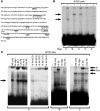
Characterization of Ikaros-type transcription factor-binding elements in the POMC gene promoter. (A) Potential Ikaros-type binding sites in the POMC –543 promoter were determined with TRANSFAC software. Five potential sites, indicated by A, B, C, D and E, were identified. Other recognized transcription factor–binding elements are indicated. (B) The 5 POMC promoter Ikaros-binding sites were used as EMSA probes with nuclear extracts from AtT20 cells. Note complex formation with fragments A, B, and C but not D and E. Reactions without (–) and with (+) nuclear protein are indicated. (C) Oligonucleotides A, B, and C were further tested in the absence or presence of 100 M excess Ikaros oligonucleotide or a monoclonal antibody against Ikaros (Ik Ab 1), a polyclonal antiserum against Ikaros (Ik Ab 2), Eos (Eos Ab), and a control antibody (Ctrl Ab) against Rb as indicated. As noted in B, the site A complex runs faster than those generated by B or C. The supershifted bands with the antibody against Ikaros are uniformly more intense and migrate slightly higher than those with the antibody against Eos. Increased amounts (μl) of Ikaros antiserum (second panel) result in increased intensity of the supershifted bands.
Figure 3
Figure 3
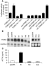
Ikaros regulation of the pituitary POMC gene. (A) The effect of Ikaros on POMC promoter activity was tested in a luciferase (luc) assay. Note activation by wild-type Ik1 and loss of activity by the Ikaros dominant negative isoform (Ik6) and by Eos. The functional role of the putative Ikaros-binding domains at sites A, B, and C in the POMC promoter was also examined. Mutation of 2 Ikaros-binding sites at sites A (mA) and B (mB) resulted in loss of reporter activity in transfected corticotroph AtT20 cells. This effect was not seen with mutation of site C (mC). (B) The effect of Ikaros or Eos transfection on endogenous POMC gene mRNA expression. AtT20 cells stably transfected with Ik1, Ik6, or Eos were examined by Northern blotting using a cDNA probe for the POMC gene. Densitometric analysis from multiple independent clones revealed an approximately 2.5-fold increase in POMC/GAPDH expression in Ik1-transfected clones. In contrast, forced expression of Eos (right) reduces POMC expression. pcDNA, cells transfected with empty vector. (C) Conditioned culture media from Ikaros-transfected cells were examined for ACTH content by ELISA. Note significantly enhanced ACTH secretion by Ikaros-transfected cells compared with empty vector in contrast to mild reduction by Ik6 or Eos-transfected cells.
Figure 4
Figure 4
Loss of Ikaros impairs pituitary corticomelanotroph development and ACTH expression. (A) The pituitaries of Ikaros-null mice (KO) at E17.5 were compared with age-matched controls from heterozygous (HET) and wild-type littermates as indicated. The sections were taken from the midline of the gland and include the sphenoid bone (S) in the midline below the gland. Note the normal onset and distribution of the corticomelanotroph-specific Tpit factor (upper panels) but reduced ACTH reactivity (lower panels) in heterozygous mice and near complete absence of ACTH in homozygous mice. (B) By 1 week of age, immunocytochemical examination reveals persistent loss of ACTH reactivity in the pituitary of Ikaros-null mice compared with wild-type control littermates. The heterozygous (HET) animals also showed loss of ACTH immunoreactivity, but the glands were larger than those of homozygous animals, and the striking loss involved the intermediate lobe (arrows) that is separated from the anterior lobe by the residual Rathke’s cleft (*) in the mid-portion of the gland. Original magnification, ×63.
Figure 5
Figure 5
Disrupted corticomelanotroph development in Ikaros-deficient mice impairs pituitary-adrenocortical function. Levels of circulating hormones of the pituitary-adrenal axis including the POMC-derived peptides ACTH (A) and MSH (B) and the ACTH-dependent adrenocortical hormone corticosterone (C) were measured by immunoassay in Ikaros-null (n = 8), heterozygous (n = 31), and wild-type littermates (n = 18) at 3 weeks of age. The reductions in ACTH, MSH, and corticosterone levels were most pronounced in Ikaros-null mice, as illustrated by the horizontal-bar comparisons. (D) Neither lymphocyte reconstitution nor treatment with vehicle changed circulating corticosterone levels in Ikaros-deficient animals. Data are from animals described in Table 1 and reflect the impact of reconstitution with wild-type marrow cells 5 weeks after intrahepatic injection.
Figure 6
Figure 6
Disrupted pituitary-adrenocortical function in Ikaros-deficient mice contributes to impaired growth and diminished survival. (A) Health status and weight were recorded daily. Compared with Ikaros heterozygous and wild-type control littermates, Ikaros-null mice exhibit growth retardation from birth. Treatment with glucocorticoid hormone (+) compared with vehicle alone resulted in significant weight increase in Ikaros-null mice, an effect not as evident in heterozygous animals. No significant impact of glucocorticoid treatment was observed in wild-type control littermates. Values shown represent the mean weights in grams and, where statistically significant (P < 0.02), treatment effects are denoted by an asterisk. (B) Kaplan-Meier curve depicting the diminished survival of homozygous Ikaros-deficient mice treated with vehicle alone (dashed line; n = 8). The increased mortality was reversed with complete survival in hormone-treated Ikaros-null mice beyond 6 weeks as indicated by the solid line (n = 7). #P < 0.05.

Comment in

References

    1. Chrousos GP. The hypothalamic-pituitary-adrenal axis and immune-mediated inflammation. N. Engl. J. Med. 1995;332:1351–1362. - PubMed
    1. Turnbull AV, Rivier CL. Regulation of the hypothalamic-pituitary-adrenal axis by cytokines: actions and mechanisms of action. Physiol. Rev. 1999;79:1–71. - PubMed
    1. Asa SL, Ezzat S. The cytogenesis and pathogenesis of pituitary adenomas. Endocr. Rev. 1998;19:798–827. - PubMed
    1. Asa SL, Ezzat S. Molecular determinants of pituitary cytodifferentiation. Pituitary. 1999;1:159–168. - PubMed
    1. Molnar A, et al. The Ikaros gene encodes a family of lymphocyte-restricted zinc finger DNA binding proteins, highly conserved in human and mouse. J. Immunol. 1996;156:585–592. - PubMed

Publication types

MeSH terms