

Bcl-2-related protein A1 is an endogenous and cytokine-stimulated mediator of cytoprotection in hyperoxic acute lung injury
- PMID: 15841185
- PMCID: PMC1070412
- DOI: 10.1172/JCI23004
Bcl-2-related protein A1 is an endogenous and cytokine-stimulated mediator of cytoprotection in hyperoxic acute lung injury
Abstract
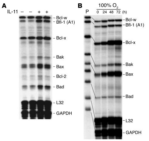
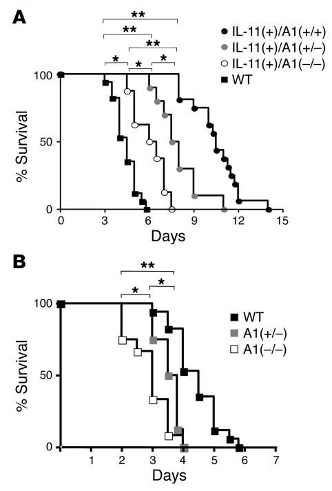
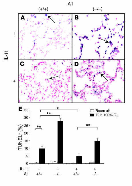
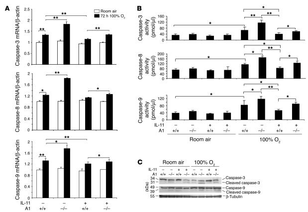
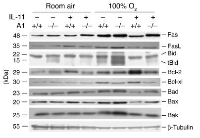
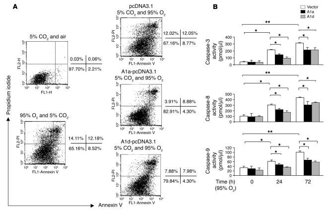
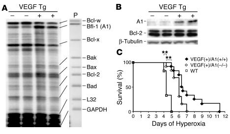
Hyperoxic acute lung injury (HALI) is characterized by a cell death response with features of apoptosis and necrosis that is inhibited by IL-11 and other interventions. We hypothesized that Bfl-1/A1, an antiapoptotic Bcl-2 protein, is a critical regulator of HALI and a mediator of IL-11-induced cytoprotection. To test this, we characterized the expression of A1 and the oxygen susceptibility of WT and IL-11 Tg(+) mice with normal and null A1 loci. In WT mice, 100% O(2) caused TUNEL(+) cell death, induction and activation of intrinsic and mitochondrial-death pathways, and alveolar protein leak. Bcl-2 and Bcl-xl were also induced as an apparent protective response. A1 was induced in hyperoxia, and in A1-null mice, the toxic effects of hyperoxia were exaggerated, Bcl-2 and Bcl-xl were not induced, and premature death was seen. In contrast, IL-11 stimulated A1, diminished the toxic effects of hyperoxia, stimulated Bcl-2 and Bcl-xl, and enhanced murine survival in 100% O(2). In A1-null mice, IL-11-induced protection, survival advantage, and Bcl-2 and Bcl-xl induction were significantly decreased. VEGF also conferred protection via an A1-dependent mechanism. In vitro hyperoxia also stimulated A1, and A1 overexpression inhibited oxidant-induced epithelial cell apoptosis and necrosis. A1 is an important regulator of oxidant-induced lung injury, apoptosis, necrosis, and Bcl-2 and Bcl-xl gene expression and a critical mediator of IL-11- and VEGF-induced cytoprotection.
Figures

Comment in
-
To live or die: a critical decision for the lung.J Clin Invest. 2005 Apr;115(4):828-30. doi: 10.1172/JCI24681. J Clin Invest. 2005. PMID: 15841170 Free PMC article.
Similar articles
-
Interleukin-6-induced protection in hyperoxic acute lung injury.Am J Respir Cell Mol Biol. 2000 May;22(5):535-42. doi: 10.1165/ajrcmb.22.5.3808. Am J Respir Cell Mol Biol. 2000. PMID: 10783124
-
IL-6 cytoprotection in hyperoxic acute lung injury occurs via suppressor of cytokine signaling-1-induced apoptosis signal-regulating kinase-1 degradation.Am J Respir Cell Mol Biol. 2009 Mar;40(3):314-24. doi: 10.1165/rcmb.2007-0287OC. Epub 2008 Sep 5. Am J Respir Cell Mol Biol. 2009. PMID: 18776134 Free PMC article.
-
IL-6 protects against hyperoxia-induced mitochondrial damage via Bcl-2-induced Bak interactions with mitofusins.Am J Respir Cell Mol Biol. 2009 Oct;41(4):385-96. doi: 10.1165/rcmb.2008-0302OC. Epub 2009 Jan 23. Am J Respir Cell Mol Biol. 2009. PMID: 19168699 Free PMC article.
-
Cytokines in tolerance to hyperoxia-induced injury in the developing and adult lung.Free Radic Biol Med. 2006 Jul 1;41(1):4-18. doi: 10.1016/j.freeradbiomed.2006.01.027. Epub 2006 Feb 17. Free Radic Biol Med. 2006. PMID: 16781448 Review.
-
Alveolar cell death in hyperoxia-induced lung injury.Ann N Y Acad Sci. 2003 Dec;1010:405-16. doi: 10.1196/annals.1299.074. Ann N Y Acad Sci. 2003. PMID: 15033761 Review.
Cited by
-
NLRP3 deletion protects from hyperoxia-induced acute lung injury.Am J Physiol Cell Physiol. 2013 Jul 15;305(2):C182-9. doi: 10.1152/ajpcell.00086.2013. Epub 2013 May 1. Am J Physiol Cell Physiol. 2013. PMID: 23636457 Free PMC article.
-
Acute lung injury and the acute respiratory distress syndrome: four decades of inquiry into pathogenesis and rational management.Am J Respir Cell Mol Biol. 2005 Oct;33(4):319-27. doi: 10.1165/rcmb.F305. Am J Respir Cell Mol Biol. 2005. PMID: 16172252 Free PMC article. Review. No abstract available.
-
Epithelial cell death is an important contributor to oxidant-mediated acute lung injury.Am J Respir Crit Care Med. 2011 Apr 15;183(8):1043-54. doi: 10.1164/rccm.201002-0181OC. Epub 2010 Oct 19. Am J Respir Crit Care Med. 2011. PMID: 20959557 Free PMC article.
-
Hyperoxic acute lung injury and ventilator-induced/associated lung injury: new insights into intracellular signaling pathways.Crit Care. 2007;11(2):126. doi: 10.1186/cc5733. Crit Care. 2007. PMID: 17466082 Free PMC article.
-
Bcl-X(L) is the primary mediator of p21 protection against hyperoxia-induced cell death.Exp Lung Res. 2011 Mar;37(2):82-91. doi: 10.3109/01902148.2010.521617. Epub 2010 Dec 4. Exp Lung Res. 2011. PMID: 21128858 Free PMC article.
References
-
- Crapo JD. Morphologic changes in pulmonary oxygen toxicity. Annu. Rev. Physiol. 1986;48:721–731. - PubMed
-
- Barazzone C, Horowitz S, Donati YR, Rodriguez I, Piguet PF. Oxygen toxicity in mouse lung: pathways to cell death. Am. J. Respir. Cell Mol. Biol. 1998;19:573–581. - PubMed
-
- Barazzone C, White CW. Mechanisms of cell injury and death in hypoxia: role of cytokines and Bcl-2 family proteins. Am. J. Respir. Cell Mol. Biol. 2000;22:517–519. - PubMed
-
- O’Reilly MA, et al. Bcl-2 family gene expression during severe hyperoxia induced lung injury. Lab. Invest. 2000;80:1845–1854. - PubMed
-
- Ward NS, et al. Interleukin-6-induced protection in hyperoxic acute lung injury. Am. J. Respir. Cell Mol. Biol. 2000;22:535–542. - PubMed
Publication types
MeSH terms
Substances
Grants and funding
LinkOut - more resources
Full Text Sources
Medical
Molecular Biology Databases
Research Materials
Miscellaneous
