. 2005 May;115(5):1221-31.
doi: 10.1172/JCI21968. Epub 2005 Apr 14.

Oxidant stress from nitric oxide synthase-3 uncoupling stimulates cardiac pathologic remodeling from chronic pressure load

Affiliations

Oxidant stress from nitric oxide synthase-3 uncoupling stimulates cardiac pathologic remodeling from chronic pressure load

Eiki Takimoto et al. J Clin Invest. 2005 May.

Abstract

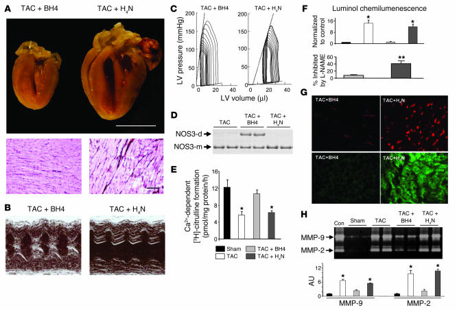
Cardiac pressure load stimulates hypertrophy, often leading to chamber dilation and dysfunction. ROS contribute to this process. Here we show that uncoupling of nitric oxide synthase-3 (NOS3) plays a major role in pressure load-induced myocardial ROS and consequent chamber remodeling/hypertrophy. Chronic transverse aortic constriction (TAC; for 3 and 9 weeks) in control mice induced marked cardiac hypertrophy, dilation, and dysfunction. Mice lacking NOS3 displayed modest and concentric hypertrophy to TAC with preserved function. NOS3(-/-) TAC hearts developed less fibrosis, myocyte hypertrophy, and fetal gene re-expression (B-natriuretic peptide and alpha-skeletal actin). ROS, nitrotyrosine, and gelatinase (MMP-2 and MMP-9) zymogen activity markedly increased in control TAC, but not in NOS3(-/-) TAC, hearts. TAC induced NOS3 uncoupling in the heart, reflected by reduced NOS3 dimer and tetrahydrobiopterin (BH4), increased NOS3-dependent generation of ROS, and lowered Ca(2+)-dependent NOS activity. Cotreatment with BH4 prevented NOS3 uncoupling and inhibited ROS, resulting in concentric nondilated hypertrophy. Mice given the antioxidant tetrahydroneopterin as a control did not display changes in TAC response. Thus, pressure overload triggers NOS3 uncoupling as a prominent source of myocardial ROS that contribute to dilatory remodeling and cardiac dysfunction. Reversal of this process by BH4 suggests a potential treatment to ameliorate the pathophysiology of chronic pressure-induced hypertrophy.


Figures

Figure 1
Figure 1
Lack of NOS3 ameliorates cardiac hypertrophy and dilatory remodeling in response to TAC-induced pressure overload. (A) Formalin-fixed (10%) hearts showing progressive cardiac hypertrophy with marked dilatory remodeling in WT TAC mice versus more modest and concentric cardiac hypertrophy at 3 weeks, with minimal further progression at 9 weeks, in NOS3–/– TAC mice. Scale bar: 10 mm. (B) Mean data for HW/TL ratio (n = 6 or more per group). (C) Histological analysis of WT and NOS3–/– TAC hearts. PAS methenamine staining reveals increased interstitial fibrosis (dark stain, upper right panel) and myocyte size in WT TAC hearts. NOS3–/– TAC hearts reveal minimal fibrosis and blunted increase in myocyte size. Scale bar: 100 μm. (D) Summary quantification of cardiomyocyte diameter (n = 4–5 per genotype, 6–10 regions per heart, 50–60 cells per heart for size estimates). P values shown indicate the effect of genotype on the TAC-stimulated response (2–way ANOVA).
Figure 2
Figure 2
In vivo hemodynamics in WT and NOS3–/– hearts subjected to TAC. (A) Representative PV loops and end-systolic and end-diastolic relations (dashed lines). In WT TAC hearts, the PV relations shifted rightward modestly at 3 weeks and markedly at 9 weeks, whereas the opposite occurred in NOS3–/– TAC hearts. LV systolic pressures at 3 weeks were similarly increased. The end-systolic PV relation (upper left relation) was steeper in NOS3–/– TAC than in WT TAC hearts. Comprehensive analysis is provided in Table 1. (B) M-mode echocardiography in conscious animals demonstrating dilated hypertrophy with decreased FS in WT TAC but concentric hypertrophy with preserved shortening in NOS3–/– TAC hearts. (C) Summary data from echocardiography (n = 5 or more per group). Wall thickness increases similarly by TAC at 3 weeks between genotypes and, at a 9 weeks, decreases slightly in WT and remains unchanged in NOS3–/– hearts. Chamber end-diastolic dimension (EDD) and end-systolic dimension (ESD) and percent FS markedly differed between the genotypes. P values shown indicate the effect of genotype on the TAC-stimulated response (2–way ANOVA).
Figure 3
Figure 3
Dot-blot analysis of fetal gene expression in LVs. (A) Examples and (B) summary quantification, with results normalized by GAPDH (n = 3–4 for each group). P values shown indicate the effect of genotype on the TAC-stimulated response (2–way ANOVA). *P < 0.01, P = 0.06 versus sham control of the same genotype.
Figure 4
Figure 4
ROS levels in WT and NOS3–/– hearts subjected to TAC. (A) Luminol chemiluminescence assay for superoxide in myocardial tissue extracts. TAC stimulated O2 formation in WT hearts, but far less in NOS3–/– hearts. P value shown reflects the effect of genotype on the TAC-stimulated response (2–way ANOVA). (B and C) Intracellular ROS generation was also estimated by red DHE staining (B) and green DCF staining (C) in frozen sections imaged by confocal fluorescent microscopy. Both signals were increased in WT TAC and strongly attenuated in NOS3–/– TAC hearts. (D) NT measured by immunofluorescent staining and quantified by ELISA assay. Both methods revealed a marked increase in NT in WT TAC hearts but low levels in NOS3–/– TAC hearts, as shown in controls for both genotypes. *P < 0.05 versus other groups. Scale bars: 50 μm.
Figure 5
Figure 5
GSH/GSSH levels, MMPs, and Akt activation. (A) HPLC determination of GSH/GSSH, xanthine, and NADPH. GSH/GSSH markedly declined with TAC in WT hearts but not NOS3–/– hearts. Xanthine increased in both, but somewhat more in WT, whereas NADPH declined similarly in both genotypes. *P < 0.05 versus sham hearts of the same genotype. (B) Gelatin zymography of myocardium in controls and following 3 weeks of TAC, and quantification results. Positive control (Con) bands for activated MMP-2 and MMP-9 are shown. Basal gel lysis was minimal but markedly increased in WT TAC hearts. This was not observed in NOS3–/– hearts either at base line or with TAC. *P < 0.05 versus other groups. (C) Response of total Akt (t-Akt) and p-Akt to TAC in both genotypes and quantification results as a ratio of p-Akt to total Akt (n = 3 per group). TAC induced a marked increase in levels of p-Akt and total Akt in WT hearts. In contrast, there was no change in NOS3–/– hearts. **P < 0.01 versus WT sham at 3 weeks.
Figure 6
Figure 6
Evidence of NOS3 uncoupling in WT TAC hearts. (A) In the WT sham heart, NOS3 appeared as both a dimer (NOS3-d) and a monomer (NOS3-m), with the largest fraction as a dimer. In boiled samples (control), the dimer was replaced by the monomeric form. The 3-week WT TAC heart exhibited largely the monomeric form, although total NOS3 expression assessed by Western blot (B) was not altered. (C) NOS Ca2+-dependent and -independent activity based on L-citrulline formation. Ca2+-dependent activity declined in WT TAC hearts (*P < 0.05). Low levels were also seen in NOS3–/– mutants, reflecting NOS1 activity. Ca2+-independent NOS2 activity showed little change. (D) Impact of pharmacological NOS3 inhibition on luminol chemiluminescence assay. Coincubation with 1 mM L-NAME inhibited 50% of luminol chemiluminescence in 3-week and 9-week WT TAC heart lysates and inhibited less than 15% at base line, supporting an increased role of NOS in O2 generation with TAC. Corresponding data for NOS3–/– myocardium are shown at right. *P < 0.05 versus WT sham at 3 weeks.
Figure 7
Figure 7
BH4, but not H4N, prevented NOS3 uncoupling, ROS generation, and cardiac remodeling induced by 3-week TAC. (A) Formalin-fixed (10%) hearts (upper panel) and histology (PAS methenamine; lower panel) showing concentric hypertrophy with BH4 cotreatment versus dilative hypertrophy with H4N accompanied by increased interstitial fibrosis. Scale bars: 10 mm (upper panel); 100 μm (lower panel). (B and C) Representative M-mode echocardiography (B) and PV loops (C) reveal corresponding functional improvement in BH4-treated, but not H4N-treated, hearts. (D) NOS3 dimer was preserved in BH4-treated, but not H4N-treated, hearts. (E) NOS Ca2+-dependent activity was restored by BH4 but not H4N treatment. *P < 0.05 versus sham. (F) Luminol chemiluminescence shows a decline in O2 generation in WT TAC hearts treated with BH4 but minimal effect with H4N treatment. *P < 0.05 versus sham. Lower graph shows the percent of luminol signal blunted by coincubation with L-NAME, confirming reduced NOS-derived O2 in BH4-treated hearts. **P < 0.05 versus BH4-treated. (G) Confocal images of DHE-stained (red) and DCF-stained (green) myocardium from WT TAC hearts treated with either BH4 or H4N. Scale bar: 50 μm. (H) Gelatin zymography for hearts with BH4 or H4N treatment and quantification results. The increased gel lysis in WT TAC hearts was reduced by BH4, but not H4N, therapy. *P < 0.05 versus sham.

Similar articles

Cited by

References

    1. Frey N, Olson EN. Cardiac hypertrophy: the good, the bad, and the ugly. Annu. Rev. Physiol. 2003;65:45–79. - PubMed
    1. Kenchaiah S, Pfeffer MA. Cardiac remodeling in systemic hypertension. Med. Clin. North Am. 2004;88:115–130. - PubMed
    1. Sorescu D, Griendling KK. Reactive oxygen species, mitochondria, and NAD(P)H oxidases in the development and progression of heart failure. Congest. Heart Fail. 2002;8:132–140. - PubMed
    1. Takano H, et al. Oxidative stress-induced signal transduction pathways in cardiac myocytes: involvement of ROS in heart diseases. Antioxid. Redox Signal. 2003;5:789–794. - PubMed
    1. Sawyer DB, et al. Role of oxidative stress in myocardial hypertrophy and failure. J. Mol. Cell. Cardiol. 2002;34:379–388. - PubMed

Publication types