. 2005 May;115(5):1163-76.
doi: 10.1172/JCI23424. Epub 2005 Apr 14.

Adipocyte-derived collagen VI affects early mammary tumor progression in vivo, demonstrating a critical interaction in the tumor/stroma microenvironment

Affiliations

Adipocyte-derived collagen VI affects early mammary tumor progression in vivo, demonstrating a critical interaction in the tumor/stroma microenvironment

Puneeth Iyengar et al. J Clin Invest. 2005 May.

Abstract

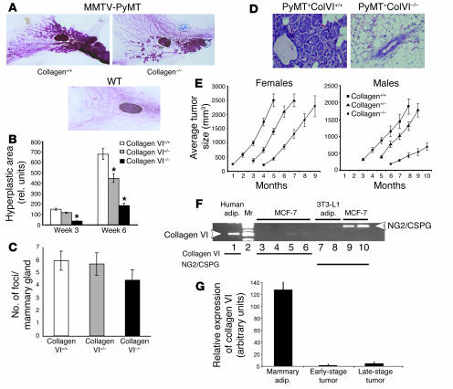
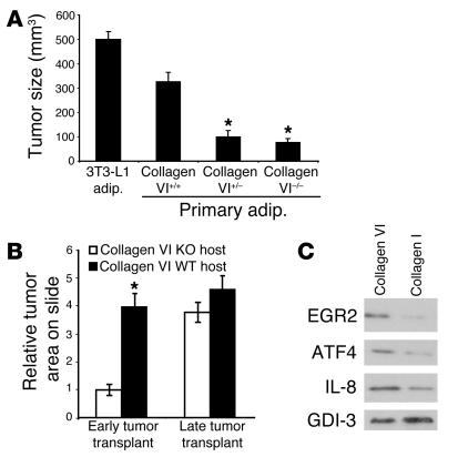
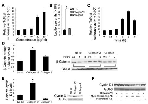
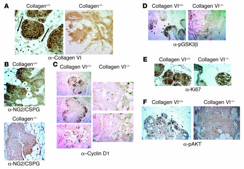
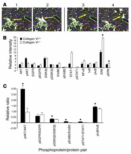
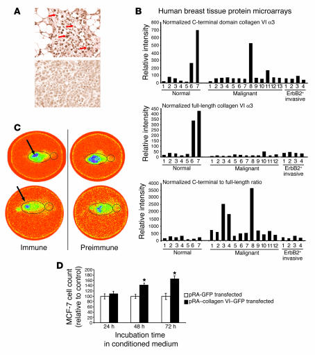
The interactions of transformed cells with the surrounding stromal cells are of importance for tumor progression and metastasis. The relevance of adipocyte-derived factors to breast cancer cell survival and growth is well established. However, it remains unknown which specific adipocyte-derived factors are most critical in this process. Collagen VI is abundantly expressed in adipocytes. Collagen(-/-) mice in the background of the mouse mammary tumor virus/polyoma virus middle T oncogene (MMTV-PyMT) mammary cancer model demonstrate dramatically reduced rates of early hyperplasia and primary tumor growth. Collagen VI promotes its growth-stimulatory and pro-survival effects in part by signaling through the NG2/chondroitin sulfate proteoglycan receptor expressed on the surface of malignant ductal epithelial cells to sequentially activate Akt and beta-catenin and stabilize cyclin D1. Levels of the carboxyterminal domain of collagen VIalpha3, a proteolytic product of the full-length molecule, are dramatically upregulated in murine and human breast cancer lesions. The same fragment exerts potent growth-stimulatory effects on MCF-7 cells in vitro. Therefore, adipocytes play a vital role in defining the ECM environment for normal and tumor-derived ductal epithelial cells and contribute significantly to tumor growth at early stages through secretion and processing of collagen VI.


Figures

Figure 1
Figure 1
Lack of collagen VI leads to a reduction in tumor growth. (A) Whole-mount analysis of early hyperplasia in 6-week-old collagen VI+/+ or collagen VI–/– MMTV-PyMT transgenic mice. Bottom panel: whole mount from a WT mouse. (B) Collagen VI reduction in hyperplastic foci size. Quantitation of hyperplastic areas at 3 and 6 weeks. n = 5 mice per group. *P < 0.05 vs. WT. (C) Number of hyperplastic foci does not significantly depend on collagen VI presence or absence in the MMTV-PyMT mouse. Quantitation of the number of foci formed in the mammary glands of 3-week-old MMTV-PyMT mice. n = 5 mice per group. (D) A representative H&E stain of a mammary section taken from PyMT+ColVI–/– and PyMT+ColVI+/+ mice at 6 weeks of age. (E) Quantitation of tumor sizes of female and male PyMT+ mice at different ages. n = 10 for each group. The 3 largest lesions were measured in each mouse. (F) Adipocytes (adip), but not breast cancer cells, express collagen VI, whereas breast cancer cells, but not adipocytes, express the collagen VI receptor NG2/CSPG. RT-PCR was performed to determine expression levels of NG2 and collagen VI. Lane 1, human adipocytes; lane 2, Marker (Mr; DNA ladder); lanes 3 and 4, MCF-7 cells; lanes 5 and 6, MCF-7 cells with adipocyte-conditioned medium; lanes 7 and 8, 3T3-L1 adipocytes; lane 9, MCF-7 cells; lane 10, MCF-7 cells with adipocyte-conditioned medium. (G) Expression of collagen VI in mammary adipocytes and early- and late-stage tumors. Quantitative real-time PCR was conducted. GAPDH was used as an internal control. Values for early-stage tumor samples were arbitrarily set to 1.
Figure 2
Figure 2
Adipocytes from collagen VI+/– and collagen VI–/– mice are less potent stimulators of tumor growth. Collagen VI also has greater impact on early than on late tumor progression. (A) SUM159-PT cells (1 × 10⁵) were coinjected into 8-week-old nude mice with the same number of isolated primary mammary adipocytes from collagen VI+/+, collagen VI+/–, collagen VI–/– mice, or 3T3-L1 adipocytes. Four weeks after injection, the sizes of the resulting foci were measured. n = 4 mice in each cohort. Results are shown as mean ± SEM. (B) MMTV-PyMT transgenic mice were sacrificed, and large, late tumor sections (from lesions larger than 1,500 mm3) and some small transformed mammary tissue for early tumor samples (lesions smaller than 300 mm3) were isolated. Tumor pieces were transplanted into partially cleared mammary glands of collagen VI+/+ and collagen VI–/– mice (n = 4). After 3 weeks, whole mounts were made. The tumor focus had a dense cellular make-up in the WT background, compared with reduced cellular density in the collagen VI–null host. Tumor areas were quantitated. Note that the absence of collagen VI protein does not affect the growth of late-stage tumor transplants. *P < 0.05. (C) Collagen VI induces proteins whose gene products are upregulated on the DNA microarrays. MCF-7 cells were treated for 6 hours with collagen VI or collagen I (each at 30 μg/ml) and extracts were generated for Western blots. EGR2, ATF4, IL-8, and GDI-3 as a loading control were measured. The levels of induction seen were similar to those seen by DNA microarrays.
Figure 3
Figure 3
Collagen VI stabilizes β-catenin and cyclin D1. (A) Dose response to increasing concentrations of collagen VI protein. A Tcf/Lef luciferase reporter construct was transfected into MCF-7 cells. Collagen VI treatment was for 3 hours. (B) Collagen VI–mediated Tcf/Lef induction. Luciferase activity was analyzed after treatment with vehicle, collagen VI (30 μg/ml), or collagen I (30 μg/ml) for 3 hours. (C) Time course of induction of Tcf/Lef activity. Luciferase activity was analyzed at different time points after treatment with 30 μg/ml collagen VI. (D) β-Catenin stability after collagen exposure. MCF-7 cells were metabolically labeled and then chased in the presence of cycloheximide for 30 minutes, 1 hour, and 2 hours. The chase medium contained vehicle, collagen VI (30 μg/ml), or collagen I (30 μg/ml). β-Catenin and GDI-3 were immunoprecipitated and quantitated. The ratio of β-catenin at 2 hours compared to 30 minutes was plotted as an indicator of β-catenin stability (3 independent experiments). No txt, no treatment. (E) Cyclin D1 stability after collagen exposure. MCF-7 cells were exposed for 3 hours to vehicle, human collagen VI (30 μg/ml), or collagen I (30 μg/ml). Cells were then lysed and analyzed for cyclin D1 by Western blot analysis. (F) Collagen VI acts in part through NG2 in MCF-7 cells to stabilize cyclin D1. In the presence of increasing amounts of NG2 neutralizing antibody in the medium (+, 0.15 μg/ml; ++, 0.45 μg/ml; +++, 0.75 μg/ml; ++++, 1 μg/ml), cyclin D1 levels decrease in a dose-dependent manner. Results are shown as mean ± SEM. *P < 0.05.
Figure 4
Figure 4
Immunohistochemical analysis of pathologically matched tumors from collagen+/+ and collagen–/– mice in the MMTV-PyMT background. (A) Absence of collagen VIα3 signal in collagen–/– mice. Staining for collagen VIα3 (with carboxyterminal polyclonal antibody) on mammary sections from pathologically matched hyperplasias developed in collagen+/+ and collagen–/– mice. Arrows point in the direction of increased levels of staining. Asterisks denote local mammary adipocytes. Magnification, ×25. (B) NG2/CSPG is expressed at the highest levels in malignant regions adjacent to adipocytes in WT mice. Note the positive staining in WT backgrounds on the leading edges of the tumor mass. Magnification, ×25. (C) Cyclin D1 is expressed at the highest levels in malignant regions adjacent to adipocytes. Magnification, ×10. (D) GSK3β is phosphorylated at the highest levels in malignant regions adjacent to adipocytes. Magnification, ×10. (E) Ki67 expression is present in cells proliferating throughout the mammary lesions. Staining for the proliferation marker Ki67 in pathologically matched lesions from WT and collagen VI–null mice demonstrates diffuse expression throughout the tumor masses in both genotypes. Magnification, ×25. (F) Akt is phosphorylated and activated at higher levels in tumors that have developed in the presence of collagen VI signaling. Staining for the ser473-phosphorylated Akt in pathologically matched tumors from collagen VI WT and knockout mice demonstrated significant staining in cells on the tumor periphery in the WT mice but not in collagen VI–null mice. Asterisks denote local mammary adipocytes in close proximity to the malignancies. Arrowheads point to the pAkt-positive cells. Magnification, ×25.
Figure 5
Figure 5
Breast tumor cell protein expression and activation differences between collagen VI+/+ and collagen VI–/– mice in the background of the MMTV-PyMT transgene. (A) Laser-capture microdissection was used to isolate hyperplastic cells in a 100-μM radius from adipocytes. A representative example is shown in the top panels, before (panel 1) and after (panel 2) removal of cells. Similarly, cells not associated at all with adipocytes (> 500 μM from the nearest adipocyte) were isolated from pathologically matched sections from mice carrying the MMTV-PyMT transgene in either a collagen VI+/+ or a collagen VI–/– background (panels 3 and 4). Arrows indicate the targeted areas before and after isolation of cells. (B) Material from at least 8 independent areas was pooled in each case and analyzed. The material was used to make protein extracts that were spotted on protein arrays. The results shown were obtained from arrays of cells in close proximity to adipocytes. Arrays were probed with various antibodies against total and activated forms of proteins implicated in pro-oncogenic pathways. *P < 0.05. (C) Quantitative comparison of the ratio of phospho-specific protein to total protein from arrays used in B. Results are shown as mean ± SEM. *P < 0.05.
Figure 6
Figure 6
Increased levels of the C-terminal domain of collagen VIα3 in tumor cells in the proximity of adipocytes. (A) High levels of the carboxyterminal domain of collagen VIα3 in a human mammary tumor sample. Top panel: collagen VI staining around human breast carcinoma cells near the vicinity of the adipocytes (arrows). Bottom panel: reduced staining in tumor cells more distant from adipocytes in the same tumor section. (B) Polyclonal-to-monoclonal ratio of collagen VI. Reverse-phase protein arrays determined levels of the C-terminal domain from collagen VIα3 (top) and the full-length collagen VI protein (middle) found in normal and tumor tissue of human cancer specimens. Bottom panel: ratio of relative signals obtained for polyclonal and monoclonal antibodies, indicating a bias toward higher levels of the α3 C-terminal domain. (C) The carboxyterminal fragment of collagen VIα3 accumulates on the surface of tumors. Polyclonal IgGs from immune and nonimmune preparations against the carboxyterminal domain of collagen VIα3 were radiolabeled with 188-rhenium. Preparations were injected either i.p. (top) or i.v. in the tail vein (bottom) into 10-week-old MMTV-PyMT mice. The outline of the mouse is indicated. Mice were imaged on a Siemens LEM+ZLC DIGITRAC gamma camera. (D) Recombinant carboxyterminal C3–C5 domains of collagen VIα3 display potent pro-mitogenic activity. MCF-7 cells were treated with conditioned medium containing recombinant carboxyterminal collagen VIα3 protein or conditioned medium from control transfected cells for 24, 48, or 72 hours. Cell number was normalized to the control cells at the respective time points. *P < 0.05.

Similar articles

Cited by

References

    1. Elenbaas B, Weinberg RA. Heterotypic signaling between epithelial tumor cells and fibroblasts in carcinoma formation. Exp. Cell Res. 2001;264:169–184. - PubMed
    1. Wiseman BS, Werb Z. Stromal effects on mammary gland development and breast cancer. Science. 2002;296:1046–1049. - PMC - PubMed
    1. Klausner RD. The fabric of cancer cell biology: weaving together the strands. Cancer Cell. 2002;1:3–10. - PubMed
    1. Iyengar P, et al. Adipocyte secreted factors synergistically promote mammary tumorigenesis through induction of anti-apoptotic transcriptional programs and proto-oncogene stabilization. Oncogene. 2003;22:6408–6423. - PubMed
    1. Lin EY, Nguyen AV, Russell RG, Pollard JW. Colony-stimulating factor 1 promotes progression of mammary tumors to malignancy. J. Exp. Med. 2001;193:727–740. - PMC - PubMed

Publication types